Eyelash and Corneal Ulcers in Bulldogs and French Bulldogs
Distichiasis, Distichiae, Trichiasis, and Ectopic Cilia are some of the common eyelash problems in bulldogs. The problematic eyelash can either arise from an abnormal location on the eyelid or grow in an abnormal manner.
Eyelash and Corneal Ulcers in Bulldogs and French Bulldogs ABNORMALITIES
BULLDOG TRICHIASIS
Bulldog and French Bulldogs Trichiasis, is an eyelash that grows from the right location, but in an abnormal orientation. Instead of growing away from the eyeball, it grows toward the eyeball (cornea).
Trichiasis: NORMAL LOCATION ABNORMAL DIRECTION
BULLDOG ECTOPIC CILIA
Somewhat similar to distichiasis, the bully ectopic eyelash emerged from an abnormal part of the eyelid but even further into the inside, near the conjunctiva. The bulldog’s ectopic eyelash is usually abnormally stiff, pointy, and sometimes curled.
Ectopic Cilia: ABNORMAL LOCATIN, ABNORMAL EYELASH
BULLDOG DISTICHIASIS

Somewhat similar to ectopic cilia the bulldog Distichia is an abnormal eyelash(s) that grows from an abnormal location. In contrast to normal eyelashes that grow from the outer part of the eyelid, Distichiae grows from the inner part of the eyelid and usually rises from the eyelid meibomian gland.
Distichiasis: ABNORMAL LOCATIN, ABNORMAL EYELASH
Eyelash and Corneal Ulcers in Bulldogs and French Bulldogs MEIBOMIAN GLANDS:
Your bulldog and French bulldog eyelid meibomian are small, tiny glands embedded in your bulldog eyelid. The glands secrete sebaceous material that is part of your bulldog tear film.
Bulldog and French Bulldog KCS: Your bulldog tear film has 3 parts, a water-like part produced by the tear glands and the lipid mucoid part made by the Meibomian glands. Deficiency of the lipid, mucoid, and/or aqueous part of your bulldog tear film will likely lead to the common “bulldog dry eye” (KCS)
MEIBOMIAN TUMORS: Bulldog meibomian tumors usually present as swollen bumpy glands, and are more common in older bulldogs. Most of them are benign and slow to grow (Adenomas). Nevertheless, they should be removed when they start aggravating the cornea, become sizable, or bleed.
Eyelash and Corneal Ulcers in Bulldogs and French Bulldogs DISTICHIASIS
- There is often more than one bulldog distichiae growing across the eyelid
- More than one bulldog distichia can be found in the same meibomian gland
- Distichiae is most often found in the upper lid but it can also be found growing from the meibomian gland of the lower lid.
- Bulldogs Distichiae can be found simultaneously in their bottom and upper eyelids and on their left and right eyelids
The reason why the eyelash follicles develop in this abnormal location is not known
Eyelash and Corneal Ulcers in Bulldogs and French Bulldogs PRESENTATION
DISCHARGE: excess tears and mucus due to corneal irritation and sometimes infection

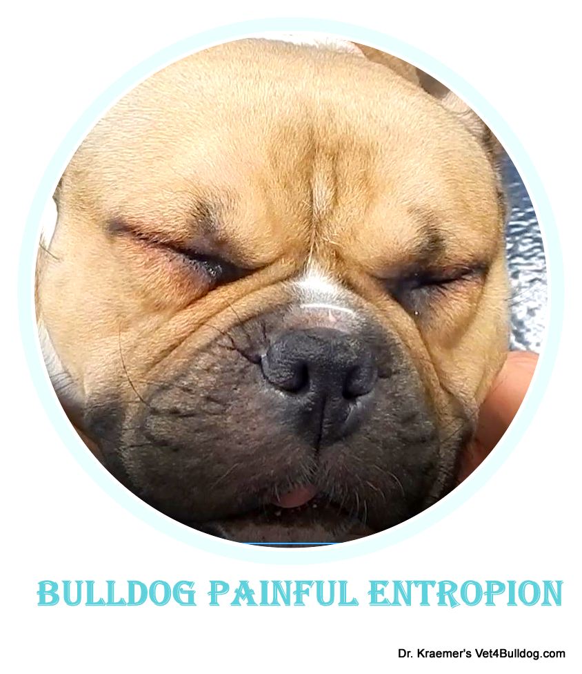
SQUINTING & RUBBING: the abnormal eyelash contact with the cornea is painful and irradiating, thus your bulldog may rub the eyes or/and squint
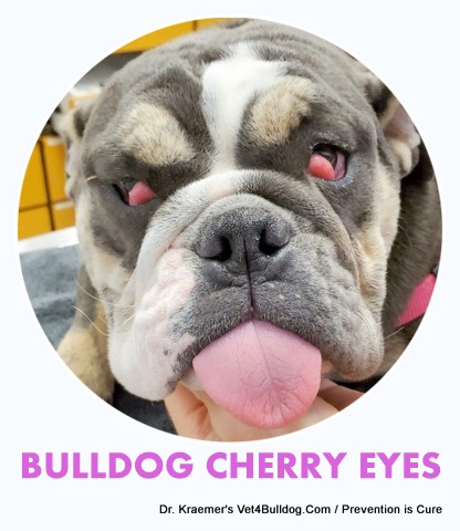
REDNESS: conjunctivitis, scleritis, and puffy eyelids
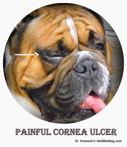
CORNEAL ULCER: corneal cloudiness, hyperpigmentation, blue or white opaque lesions, and blood vessels over the cornea might be observed.
PAIN: The intensity of the pain depends on the stiffness of the abnormal eyelashes, the number of them, and the degree of contact with the cornea
Eyelash and Corneal Ulcers in Bulldogs and French Bulldogs Diagnosis
OPHTHALMIC EXAM: a complete ophthalmic exam should be conducted.
MAGNIFICATION & ILLUMINATION: Sometimes it is relatively easy to visualize the abnormal eyelashes, other times they are colorless and very thin, thus hard to see with the naked eye
Diagnosis usually requires magnification and proper illumination.
Sedation might be required if the pet is in pain, blinking, moving his/her head or too anxious
CORNEAS STAIN: Corneal fluorescein staining should always be done to r/o an active corneal ulcer
STT: Schirmer Tear Test (STT) is also recommended to help r/o tear deficiency that might be compounding the problem. KCS is very common to bulldogs.

Eyelash and Corneal Ulcers in Bulldogs and French Bulldogs TREATMENT
WAIT & SEE: bulldogs and French bulldogs’ abnormal eyelashes might be diagnosed incidentally during a routine physical exam. The bulldog owner should be informed about it but if there is no discomfort and/or notable eyelash contact with the cornea it might be best to leave it alone. Also, in those cases, an ophthalmic lubricant can be added to serve as a barrier. Last, they can be removed and cryofreeze during an upcoming elective surgery such as spaying & neutering, a bulldog.
MANUAL PLUCKING: using fine forceps, sometimes the distichia can be plucked out
- Warning: it’s hard to do if the dog is restless and moving, in those cases, due to potential injury to the medical team and the pet the attempt should be aborted.
- Warning: Manual plucking is only a temporary solution, bully distichia is likely to reoccur, and at times the regrown hair is stiffer and worse than the original.
CRYOTHERAPY: meibomian glands can be frozen and the hair follicle(s) destroyed with a liquid nitrogen cryopen. This should help prevent new abnormal eyelashes from regrowth.
Cryotherapy requires sedation or general anesthesia. It’s the ideal treatment for bulldogs with a large number of distichia.
ELECTROLYSIS: inserting an electrode into the gland and applying an electric current could help destroy the hair follicle and prevent regrowth. This procedure is also done under anesthesia.
SURGERY: Surgery is rarely recommended and involves dissection of the gland with the hair follicle and the abnormal eyelash.
ENTROPION SURGERY: I often recommend entropion surgery that helps pull/rotate the eyelids away from the eye. This could and likely will reduce the need for repeating the distichiasis procedure in the event that there is regrowth or new distichia later on.
TOPICAL OPHTHALMIC
- LUBRICANTS
- ANTIBACTERIAL
- PAIN RELIEF

- ULCERS HEALING: usually some form of a topical hyaluronic acid ointment, serum, or PRP
PAIN RELIEF:
- NSAID
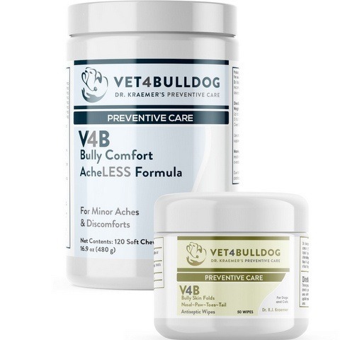
- SUPPLEMENTS: Like Dr. Kraemer’s V4b Bully Comfort & AchLess Formula Chews
Eyelash and Corneal Ulcers in Bulldogs and French Bulldogs TIPS & WARNINGS
Tip #1 E-COLLAR: keep a buster collar till the abnormal eyelash is removed and if needed also post-op to avoid rubbing and pawing at the eye.
Tip #2 TOPICAL: Topical ophthalmic medication might be needed usually just short term to reduce pain, help heal the cornea, and eliminate bacteria.
Tip #3 PAIN RELIEF RX: pain control RX/and supplements are often recommended
Tip #4 EXAM: Any time I treat a bulldog with an eye problem I examine the lids for those abnormally ingrown eyelashes. Many of those bulldog abnormal eyelashes are light-colored and thin thus easily missed.
In my exam room I always inspect bulldog’s eyelids with a magnifying head light from different angles.
Warning #1 MAGNIFYING & ILLUMINATION: Bulldog Distichiasis is difficult to see with a naked, thus I will usually use a magnifying glass and proper illumination.
Warning #2 VET: Because distichiasis is only common to certain breeds like bulldogs and French bulldogs, many veterinarians will fail to look for the problem and/or fail to see it.
Warning # 3 BREEDING; Due to the hereditary nature of those conditions bulldogs with a history of eyelash problems should be discouraged from breeding.
Warning #4 SCARING: Complications associated with electrocautery and cryosurgery may include excessive scarring of the eyelids.
Warning #5 REGROWTH & REPEAT: Regrowth of hairs may occur from the treated gland or from other glands that did not have any abnormal eyelashes at the time of the procedure, both might require a repeat of the procedure.
Warning #6 ECTOPIC CILIA: bulldog and French bulldog ectopic cilia almost always cause pain and corneal injury. They are hard to find because they are deep in the inside of the lid and very short. They should be removed surgically as soon as possible so as to avoid serious injury to the cornea
Sudden squinting, red, teary eye is often due to an emerging corneal ulcer, you should consider this problem to be an an emergency and seek help as soon as possible.

Warning #7 ENTROPION: Bulldog Entropion will often exasperate the condition and the trauma to the cornea.
Warning #8 EYE LOSS: Unattended chronic bulldog distichiasis can lead to a deep, acute, infected, melting perforated ulcer that can end with a tragic loss of your bulldog’s eye.
Warning #9 CORNEAL SCARING: Other times it can lead to chronic corneal scaring, manifested with an opaque white scar or fibrotic red one, black spots, cloudiness, and blood vessels formation over the cornea which will diminish your bully vision.
Warning #10 RUBBING & ITCHING Consideration should be given to other causes of itching and rubbing of the face, such as bulldog allergies and bulldog and French bulldog skinfold dermatitis, Those cases should be treated with allergy and itch control medication and topical as well as skinfold antiseptic bully wipes
Recommnded by Owners Approved by Bulldogs