RE: Painfull Looking Sudden Cherry Like bulldog Eye
Hello Dr. Kraemer,🩺 💉 I’m writing regarding my French Bulldog puppy, Oliver. Recently, I’ve noticed a peculiar development: his left eye seems to have a pinkish gland that pooped. It it painfull? Your guidance has been incredibly valuable to us! 🐾👀
#OliverTheFrenchie #CherryEyeConcerns ❤️ Thank you! 😊
Introduction to Cherry Eye In Bulldogs and Fr. Bulldogs
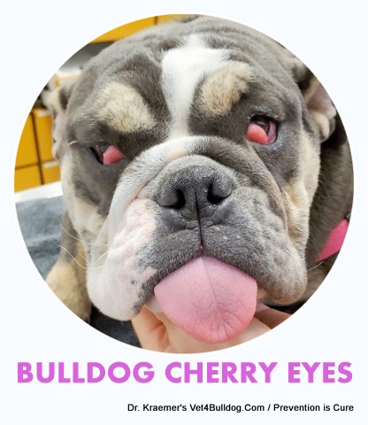
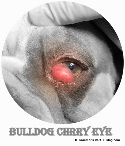
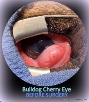
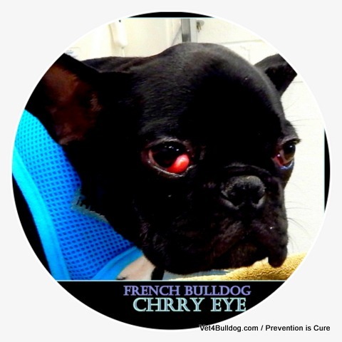
Cherry Eye in Bulldogs and French Bulldogs becomes noticeable when the gland of the third eyelid (nictitating membrane) protrudes, appearing as a pink, oval-shaped bulge. Normally, this gland is hidden and protected by the third eyelid, making it barely visible in a healthy puppy.
BULLDOG THREE EYELIDS
In contrast to us, your French Bulldogs Puppies, and your English Bulldogs Puppies, have three eyelids,
- The typical top eyelid
- The typical bottom eyelid
- A third passive pale pink-colored one originates from the nasal corner of the eyes.
Bulldog Cherry Eye 5 X MUST KNOW
- Cherry eye is a prolapse of a gland normally situated under the third eyelid gland.
- Cherry eye is common to both the English and French bulldog puppies
- % of the tear production is produced. in the cherry gland, thus it must never be removed
- When small, it can be massaged back under the lid but most times, surgery and repair will be required
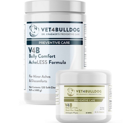
- Prevention and care include Dr. Kraemer’s Cherry Eye Bundles.
Wellocme to Our “Prevention Over RX” Bulldog Community
BULLDOG THIRD EYELID:
🚨Your bulldog’s 3ed eyelids is a pink member seen in the corner of the eyes, its medical term is “nicotinic membrane”.
The third eyelid helps to:
- PROTECT THE CORNEA: Physically protect your bulldog cornea by acting as a windshield wiper
- DISTRIBUTE TEARS: Helps distribute the tears over the eye
- SHIELD THE CHERRY: embeds, shields, and protects the tear gland (cherry)

Your bulldog puppy’s third eyelid also contains lymphoid tissue, suggesting immune protection of the eye
BULLDOG TEAR FILM 3 PARTS:
The Eye Tear Film combines three parts:
- LIPID (fat)
- MUCIN (Mucus)
- WATER (Aqueous)
- There is one just above the eye in the upper lid area
- A second one (The “Cherry Gland”) that is embedded in the third eyelid
The cherry gland produce Tears, Lubricate, hydrate, and preserve your bulldog cornea
What Are the Symptoms of Cherry Eye in Bulldogs?
- Cherry: Large, swolle,n red “cherry” protrusion at the nasal corner of the eye
- Third Eyelid: white membranous tissue adjacent to the cherry or over it
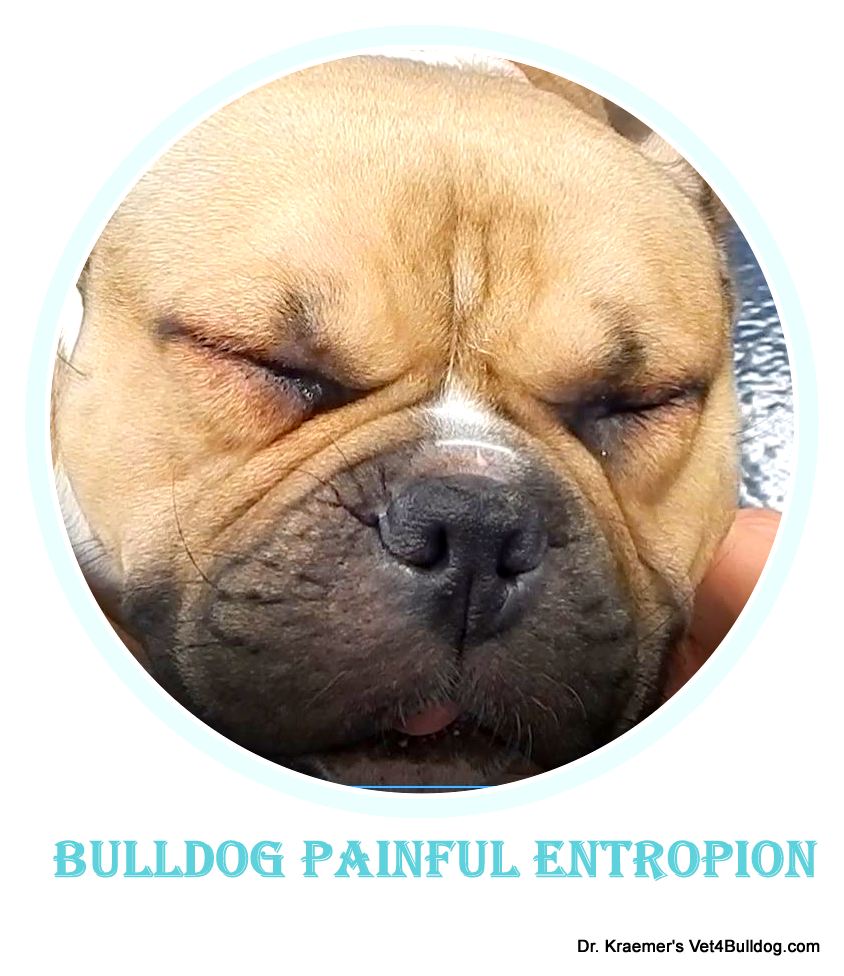
- Blink & Squint: if the cherry is rubbing and irritating the cornea
- Mucoid or Watery Discharge: If there is a corneal irritation or tear production problem
- Eye Pawing and Rubbing: If there is discomfort
What Causes the Prolapse of Bulldog Cherry Eye?
- LIGAMENTOUS WEAKNESS: weakness of the ligamentous attachments.
- FIBROUS ATTACHMENT: Weaknesses of gland fibrous attachments
- BLOCKAGE: Tear duct blockage that leads to swelling
- VASCULAZATION: Poor circulation
- INFECTION:
- conjunctivitis
- scleritis,
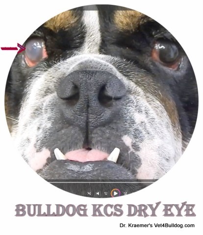
- dry eye KCS
- INFLAMMATION:
- SKULL CONFIRMATION: Skull bone confirmation (brachycephalic,) bulging eyes
- INHERITANCE: Genetics
- UNKNOWN: The “cherry gland” prolapse cause is often unknown
Cherry Eye is more common in young bulldogs puppies
How to Prevent and Treate Cherry Eye in Bulldogs and French Bulldogs?
Cherry eyes can often be massaged back into place.
You should seek veterinary advice, especially if you can’t massage it back under the lid if it’s getting worse or if there are any signs of discomfort.
In contrast to most eye problems in bulldogs, the cherry eye in its milder form is usually a non-emergency.
Cherry Eye in Bulldogs and French Bulldogs RULE OF THUMB👍
Cherry eye medical management includes
- Ophthalmic anti-inflammatory
- Ophthalmic antibiotics
- Massaging the gland back in can be attempted

MASSAGING BULLDOG CHERRY EYE
This maneuver is rarely a long-term solution, thus surgical correction is recommended.
Bulldog Cherry Eye SURGICAL REPAIR:
There are two popular cherry-eye techniques
1. CHREY EYE ANCHORING TACKING SURGERY
- ONE stitch is a single-stitch technique
- NO CUTS: There is no cutting involved
- SIMPLE: It’s more commonly done due to simplicity
- UNRELIABLE: Its holding power is limited and the chances for re-prolapsing are high
2. CHRREY EYE IMBRICATION POCKET SURGERY:
- POCKET: A “pocket” is created to help bury and secure the gland inside the third eyelid.
- CUTS: It includes cuts on the two sides of the gland and tissue removal to help create the “pocket.”
- SUTURES: The cherry is then pushed into the pocket, and the two sides are pulled tightly together over it with suture placement.
- FIBROTIC BAND: The sutured cuts will fibrously form a strong, tough band over the gland to hold it from re-prolapsing.
CHERRY EYE SURGERY & ANESTHESIA WARNING:
Before opting for surgery, you should do your due diligence, which includes
- Bulldog Anesthesia Risks
- Surgeon Qualifications and Experience
- Post-Op Complications
#1 CHERRY EYE RE-PROLAPSE WARNING:
A suture or stitch becomes undone prematurely, which can lead to an immediate re-prolapse.
#2 CHERRY EYE POST-OP ULCER WARNING:
A suture or stitch becomes undone, rubs over the cornea, and causes an injury (bulldog corneal ulcer)

#3 CHERRY EYE TACKING SURGICAL FAILURE WARNING:
The chances for failure are much higher with the tacking technique, especially with bulldogs and French bulldogs (Brachycephalic)

#4 CHERRY EYE POST-OP SWELL WARNING:
A short-term post-op swelling (edema) is likely; anti-inflammatory ophthalmic ointment might help.

Bulldog Cherry Eye Dr. Kraemer’s SURGICAL REPAIR METHOD:
For the best outcome, I combine both techniques.
- The “POCKET” provides a long-term seal
- The “TUCKING” provides a short-term anchor during the healing or swelling period
Cherry Eye in Bulldogs Dr. Kraemer’s TIPS & WARNINGS:
Below are selective cherry eye tips and warnings courtesy of Dr. Kraemer
#1 🩺CHERRY EYE SUTURE TYPE TIP:
I prefer rapidly dissolving suture material to prevent unintended suture injury to the cornea.
#2 🩺CHERRY EYE SURGICAL KNOT TIP:
The suture knot should always be placed on the outside surface of the third eyelid membrane to avoid contact with the cornea. Any corneal contact with the suture material can lead to abrasions, wounds, or corneal injury (corneal ulcers).
#3 🩺BULLDOG CHERRY EYE PREVENTION:
In many bulldogs and French bulldog puppies, only one side prolapses, yet often the other cherry eye prolapses soon after.
To prevent the other gland from prolapsing, the unaffected gland may be anchored and imbricated
#1 ⚠️CHERRY EYE OPPISTE EYE WARNING:
In many bulldogs, there’s a good chance that the opposite eye will also be “cherry.”
#2 ⚠️ CHERRY EYE RE-PROLAPSE WARNING
The risk of re-prolapse is highest with the anchor method and significantly lower when using my combination method.
Up to 30% of surgically repaired cherry eyes might re-prolapse
#3 ⚠️CHERRY EYE SURGICAL TIMING WARNING:
Cherry eye surgery in bulldogs is more likely to be successful if it’s done soon after the gland prolapses.
Over time, the prolonged chronic swelling of the gland can make the surgical repositioning more difficult, thus increasing the chance of recurrence.
#4 ⚠️CHERRY EYE SUTRE TYPE WARNING:
I don’t recommend using thick and slow-absorbing suture material. I used a very thin and rapidly absorbing suture
#5 ⚠️ CHERRY EYE PRESERVATION WARNING
Up to 30% of your bulldog’s total tear production comes from the gland in the third eyelid (the “cherry” gland). For this reason, surgical repair must prioritize preserving the gland.
Removing the “cherry” gland can significantly reduce your bulldog’s tear production, potentially leading to chronic dry eye issues.
Removal of the “cherry” will increase the chances of “bulldog dry eye” ( Bulldog Keratoconjunctivitis Sica (KCS).

#6 ⚠️ BULLDOG CHERRY REMOVAL WARNING:
Removal of the “cherry” is strictly reserved for rare cases of cancer and trauma.
#7 ⚠️ CHERRY EYE PAIN WARNING:
Post-op blinking and squinting are consistent with pain, are usually due to a corneal injury, and must be considered an emergency. Please see your vet as soon as possible.
The chances are that the suture material tore or became loose, or the knot is rubbing against the cornea and forming a corneal ulcer.
#8 ⚠️ CHERRY EYE SWELL & REDNESS WARNING:
Postoperative swelling is common following cherry eye repair, particularly with the pocket technique.
This swelling typically subsides over 2-3 weeks, during which the eye should gradually become more comfortable and regain its normal appearance.
However, if the eye suddenly becomes painful or looks abnormal, it’s important to have it rechecked by your veterinarian immediately.
Recommended by Owners Approved by Bulldogs


