


Brachycephalic dogs have reduced sensation across the surface of the cornea, which is the opposite of what you would want in a breed whose eye also sits further forward, in a shallower socket, behind a blink that does not fully close, with facial hair pointing the wrong way. Put those together and you get a dog who takes damage with far less complaint than a Labrador would make about a speck of dust.
So owners routinely find out late, and then feel terrible about it. Please do not. You were not ignoring your dog, you were reading a dog who was not giving you much to read.
There are three ways a lash causes this and one of them cannot be seen without magnification and turning the eyelid inside out, which is exactly why some dogs get their lashes plucked and ulcerate again a good few weeks later. And the test that settles whether there is an ulcer at all takes about a minute and costs almost nothing. Eyes in this breed do not give you much time, so this is the one area where I would not take a wait and see approach, and the judgment on urgency belongs with your own veterinarian.
My bulldog has had three corneal ulcers in a year. The first time our vet found extra eyelashes and plucked them and it healed beautifully. It has happened twice since. Each time we get drops and a cone and it settles, and each time I think we have fixed it. He never cries and he never paws at it, I only notice because the eye looks a bit cloudy or wet. I am starting to dread looking at him in the morning. Are we missing something?
Yes, probably, and the pattern you have described is the clue rather than bad luck.
Plucking a lash removes the shaft and leaves the follicle, so the lash grows back. That is not a criticism of your vet, it is what plucking is: a haircut rather than a cure. Which means a dog whose ulcers return on that sort of rhythm is usually a dog whose lashes have simply grown again, and the options that actually destroy the follicle are different procedures with different names.
The other thing I would want excluded is an ectopic cilium, the third of the three lash problems. That one grows through the inner surface of the lid and points directly at the cornea like a splinter, and it cannot be seen by looking at your dog in a consulting room. It needs magnification and the lid turned inside out, in a dog who is sedated or anesthetized, because no conscious Bulldog is permitting that examination. A dog with repeated ulcers in the same eye and no obvious cause deserves that look.
On him never crying: that is the part of your message I would take the most seriously, and it is not stoicism. Reduced corneal sensation is normal in this breed, so pain is a poor warning system here and cloudy or watery is often all you get. You are not missing signs, you are being given fewer of them.
What I would do with this is ask your veterinarian two specific questions: whether the lashes were plucked or the follicles were destroyed, and whether the inner lid has been examined under magnification. Those two questions tend to move a case like this along.
Dr Kraemer Oink, my 2-year-old Fr bulldog, has a painful red eye, blinking and squinting nonstop and trying to rub it. His vet tested him for dry eye 😭and ulcer but could not find anything. I need to see you ASAP🩺 ? Anything you can recommend till then?
Thank you!❤️#BulldogVet #EyeRubbing #EyePain
Introduction To Abnormal Eyelash Leading to Corneal Ulcers in Bulldogs
Distichiasis, Distichiae, Trichiasis, and Ectopic Cilia are common eyelash disorders in bulldogs.
These abnormal eyelashes may grow from an unusual location on the eyelid or in an irregular direction, often leading to irritation, pain, discomfort, and potential damage to the eye.

Most Common Abnormal Eyelash in Bulldogs and French Bulldogs
There are three common abnormal eyelashes in bulldogs
#1> BULLDOG TRICHIASIS
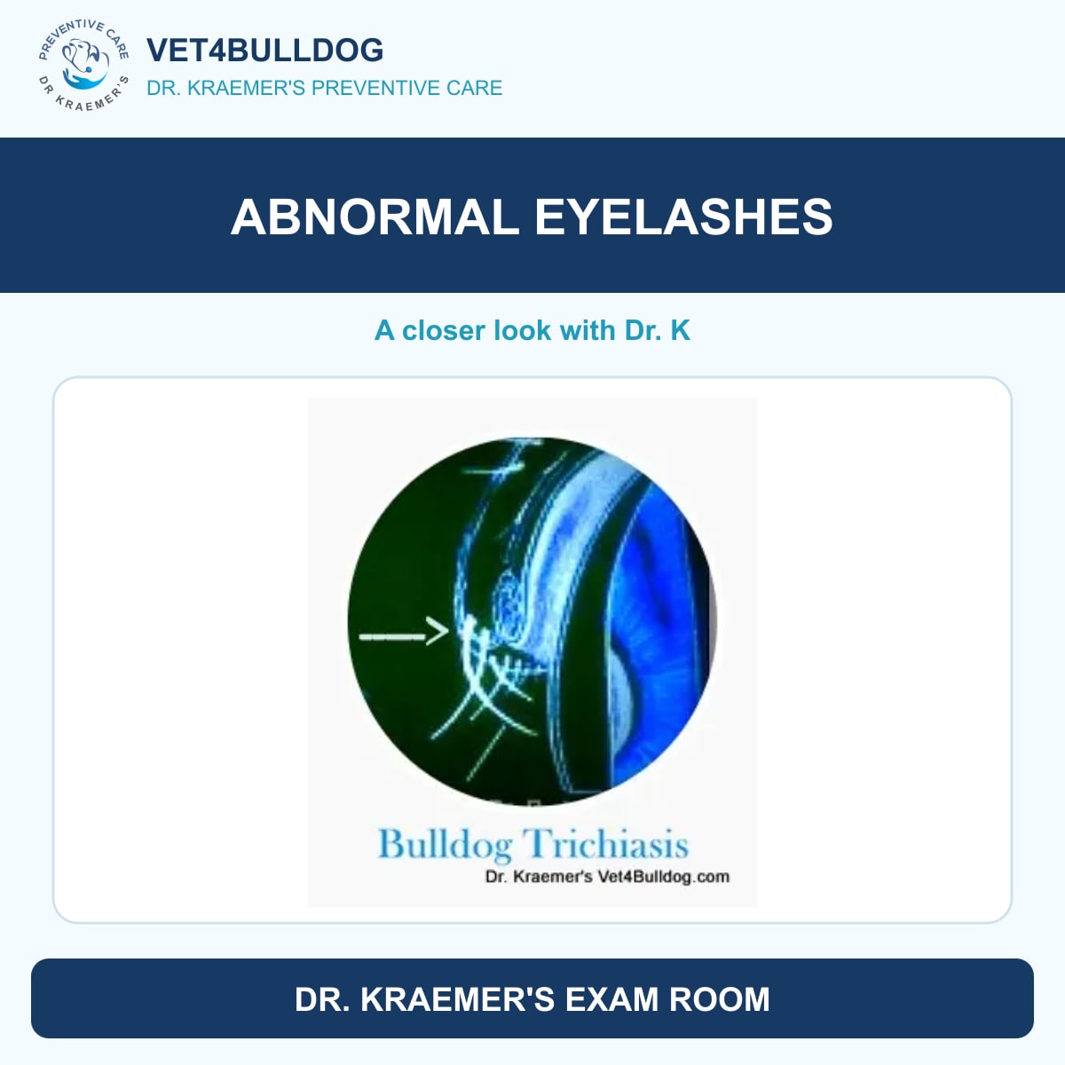
Bulldogs and French Bulldogs Trichiasis is an eyelash that grows from the right location but in an abnormal orientation. Instead of growing away from the eyeball, it grows toward the eyeball (cornea).
Trichiasis: is an eyelash gorowing from a normal eylid loction but in abnormal inward direction

#2> BULLDOG DISTICHIASIS
Distichiae in bulldogs refers to abnormal eyelashes that grow from an unusual location. Unlike normal eyelashes that emerge from the outer eyelid, distichiae grow from the inner eyelid, typically arising from the meibomian gland. These misplaced lashes can cause irritation, discomfort, and potential eye damage.
Distichiasis: eyelashes growing from an abnormal eyelid location

#3> BULLDOG ECTOPIC CILIA
Similar to distichiasis, ectopic cilia in bulldogs emerge from an abnormal location deeper inside the eyelid, near the conjunctiva. These misplaced eyelashes are typically stiff, pointy, and sometimes curled, often causing significant irritation or corneal damage.
Ectopic Cilia is an stiff pointy curled eyelash growing in an abnormal loction

Bulldog Eyelash Disorder 5 X MUST KNOW
- Eyelash disorders are unique to bulldogs, the most common of which are distchiasis, trichiasis, and ectopic cilia
- The ingrown or abnormal eyelash rubs the cornea, causing pain and potential corneal injuries
- Bulldog eyelash disorders are often missed and best diagnosed with proper light and magnification.
- Repearing them often requires cryofreezing or surgical removal
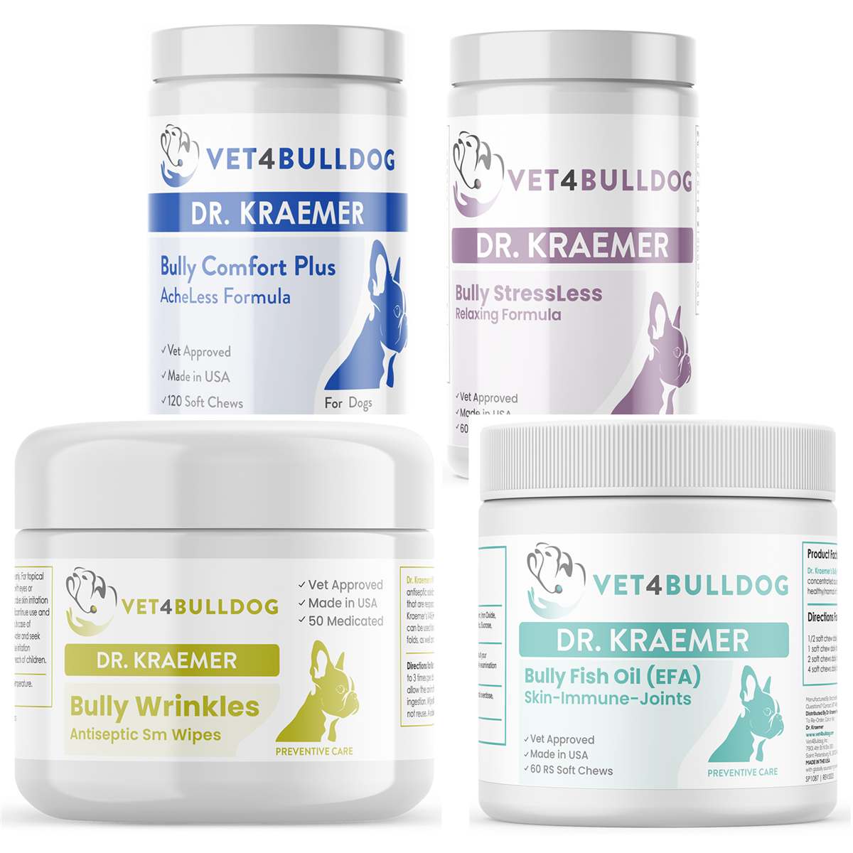
- Prevention and care include Dr. Kraemer’s Bulldog Ophthalmic Bundles.
Members of this bulldog community prefer prevention over RX

Meibomian Glands in Bulldogs and French Bulldogs
The meibomian glands are tiny sebaceous glands embedded in your bulldog’s eyelids. They secrete an oily substance that plays a crucial role in maintaining a healthy tear film.
Your bulldog’s tear film consists of three essential layers:
➡️ Aqueous layer: produced by the tear glands
➡️ Lipid (oily) layer: produced by the meibomian glands
➡️ Mucoid layer: helps stabilize the tear film
Bulldog and French Bulldog KCS (Dry Eye)
A deficiency in any of these layers, especially the lipid, mucoid, or aqueous components, can lead to bulldog dry eye (keratoconjunctivitis sicca or KCS), a common condition that affects tear production and eye health.

Meibomian Tumors in Bulldogs
Meibomian tumors in bulldogs typically appear as swollen, bumpy growths on the eyelid margins.
They are more common in older bulldogs and are usually benign adenomas, meaning they grow slowly and rarely spread. However, these tumors should be removed if they:
- Irritating or rubbing against the cornea
- Grow significantly in size
- Start bleeding or ulcerating
Bulldog Disticha and Distichiasis Must Know
🚨 There is often more than one bulldog distichia growing across the eyelid
🚨 More than one bulldog distichia can be found in the same meibomian gland
🚨 Distichiae is most often found in the upper lid, but it can also be found growing from the meibomian gland of the lower lid.
🚨 Bulldogs Distichiae can be found simultaneously in their bottom and upper eyelids and on their left and right eyelids
The reason why the eyelash follicles develop in this abnormal location is not known
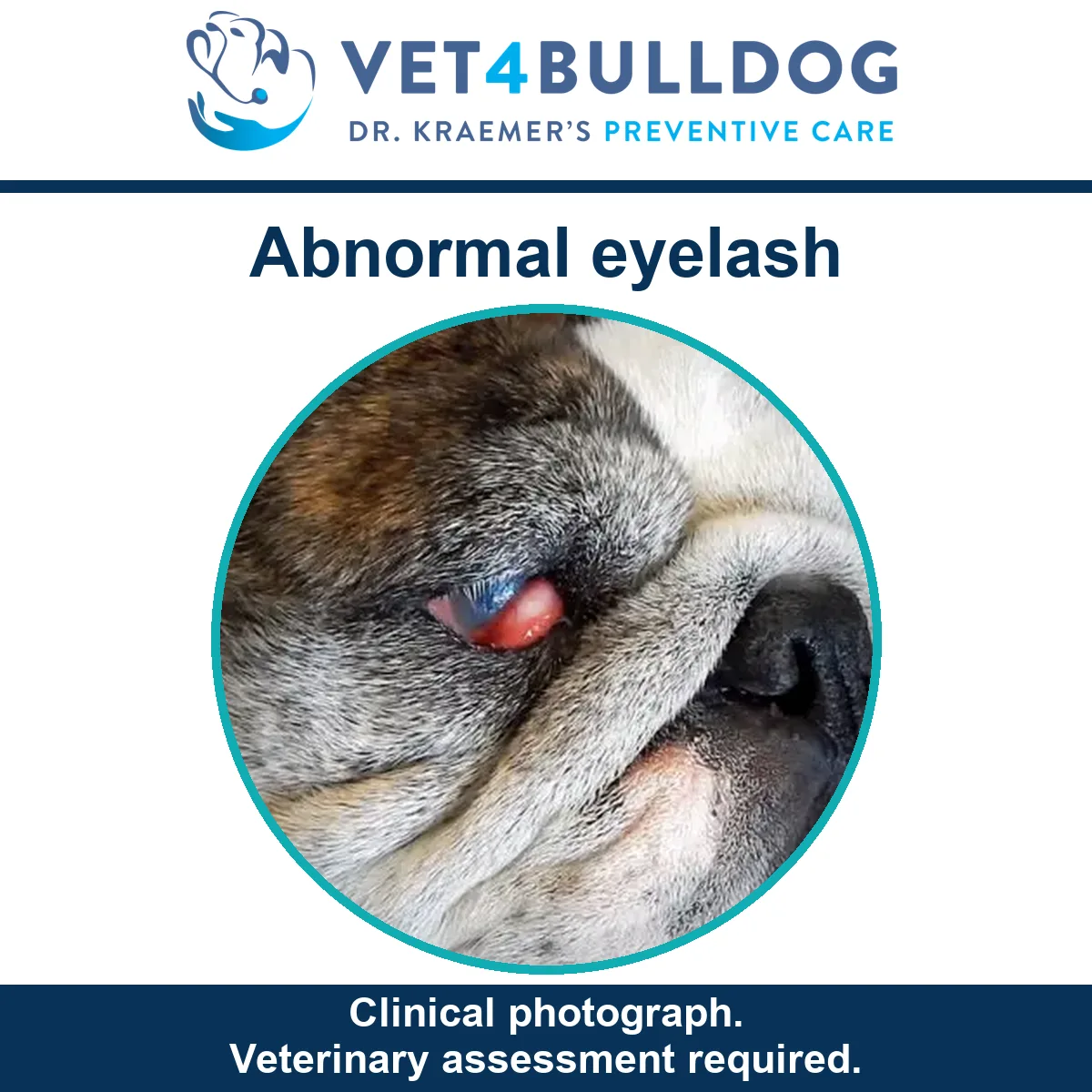
Symptoms of Abnormal Eyelashes in Bulldogs and French Bulldogs
Abnormal eyelashes in bulldogs can cause significant eye discomfort, leading to secondary complications. C
Common symptoms include:
1. Eye Discharge
- Excessive tearing and mucus production
- Often due to corneal irritation and, in some cases, infection
2. Squinting & Rubbing
- The abnormal eyelashes make painful contact with the cornea
- Bulldogs may rub their eyes frequently or squint to relieve discomfort
3. Redness & Inflammation
- Conjunctivitis (red, irritated inner eyelids)
- Scleritis (inflammation of the white part of the eye)
- Puffy, swollen eyelids
4. Corneal Ulcers & Damage
- Corneal cloudiness or hyperpigmentation
- Blue or white opaque lesions
- Blood vessels growing over the cornea (vascularization)
5. Pain & discomfort:
Pain severity depends on:
- The stiffness of the abnormal eyelashes
- The number of abnormal lashes
- The extent of corneal contact
Diagnosis of Eyelash Disorders and Corneal Ulcers in Bulldogs and French Bulldogs
The proper diagnosis of abnormal eyelashes and corneal ulcers in bulldogs requires a thorough ophthalmic examination.
1. 🩺 Ophthalmic Exam
A complete eye examination should be performed to assess the eyelids, eyelashes, and cornea.
2. 🩺 Magnification & Illumination
Some abnormal eyelashes are easily visible, while others are thin, colorless, and difficult to detect with the naked eye. Diagnosis often requires magnification and proper illumination for accurate detection.
Sedation may be needed if the bulldog is in pain, blinking excessively, moving its head, or feeling anxious.

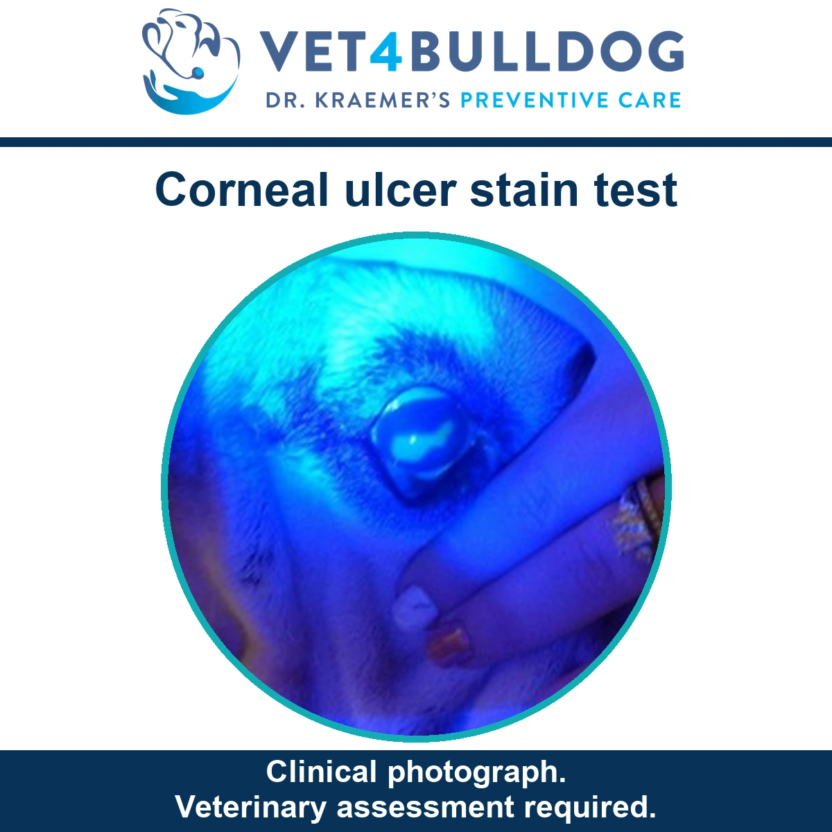
3. 🩺 Corneal Staining
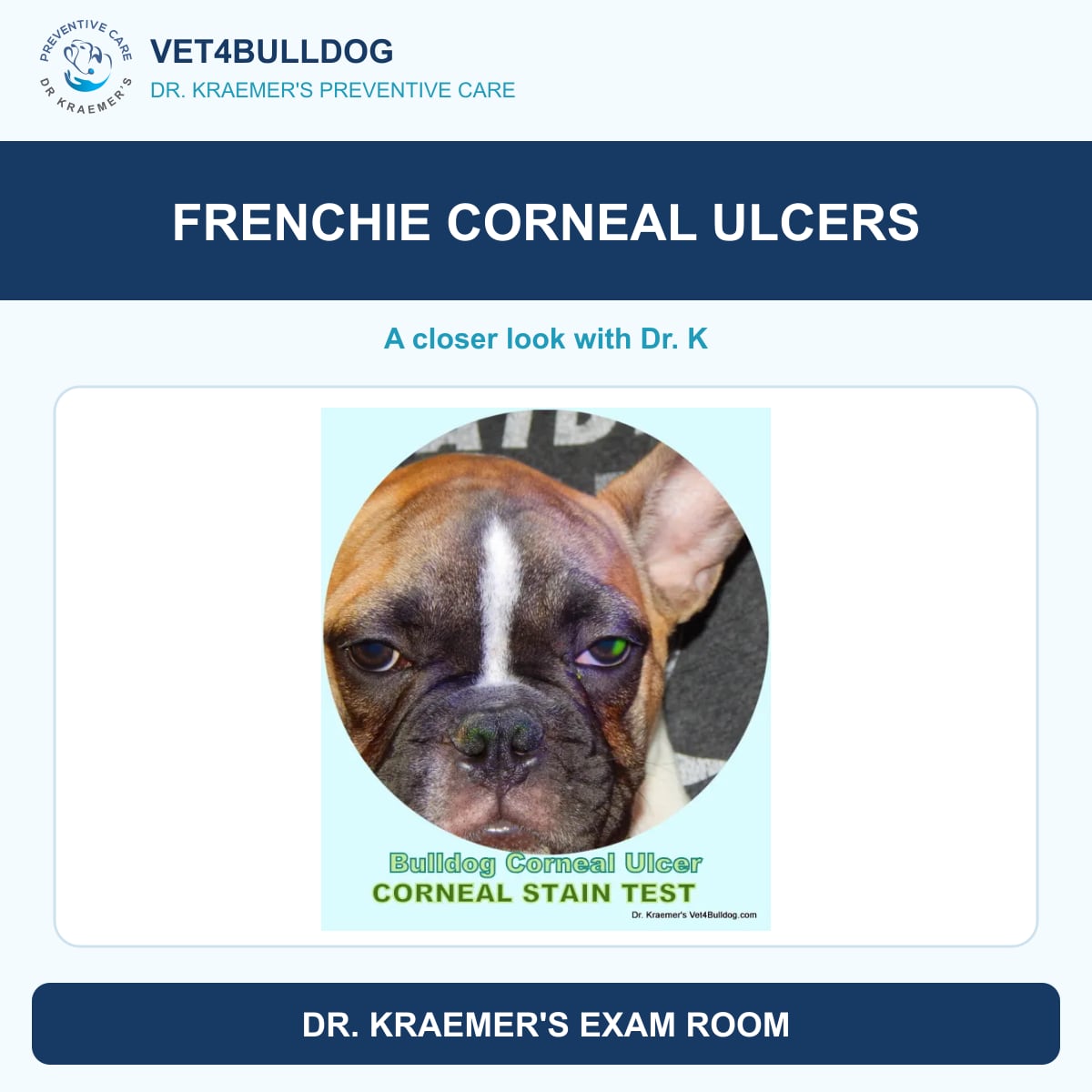
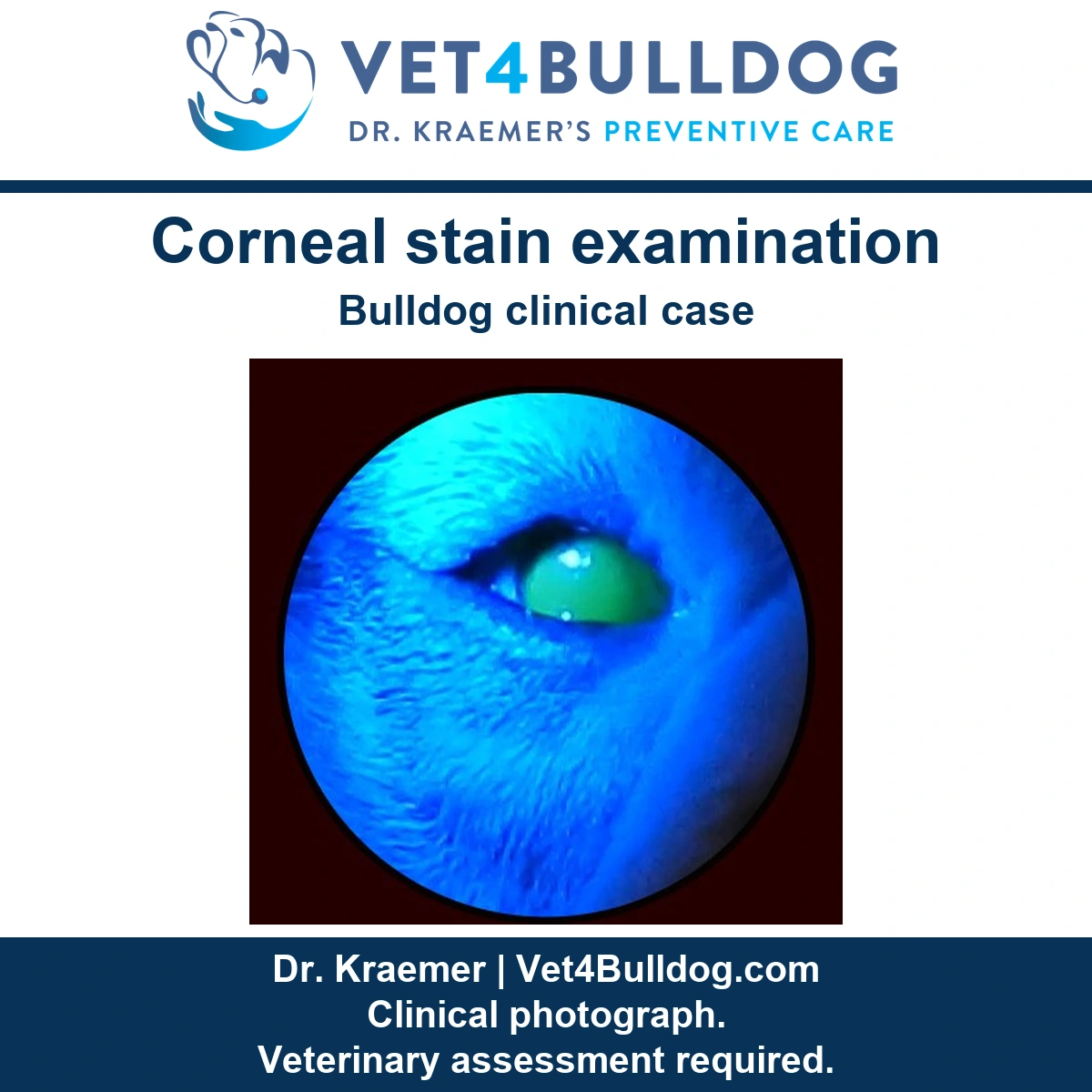
Fluorescein staining should always be performed to rule out (r/o) corneal ulcers, which are common in bulldogs with abnormal eyelashes.
4. 🩺 Schirmer Tear Test (STT)
The Schirmer Tear Test (STT) is recommended to check for tear deficiency (KCS).
Keratoconjunctivitis sicca (KCS or dry eye) is common in bulldogs and can worsen corneal damage

How to Treat Eyelash Disorders in Bulldogs and French Bulldogs?
You can take the wait & see approach
➡️ Abnormal eyelashes are sometimes detected incidentally during a routine exam.
➡️ If no discomfort or corneal contact is observed, immediate treatment may not be necessary.
➡️ Ophthalmic lubricants can be used as a protective barrier.
➡️ If elective surgery (e.g., spay/neuter) is planned, abnormal eyelashes can be removed and cryofrozen at that time.
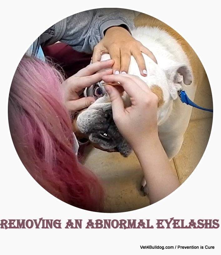
#1 Eyelash Manual Plucking
Abnormal eyelashes can be plucked out using fine forceps.
⚠️ Warnings: It can be difficult to perform on restless or anxious dogs due to potential injury.
⚠️Custion: plucking is only a temporary solution; the hair is likely to regrow, often stiffer and more irritating than before.
#2 Eyelash Cryotherapy (Cryoepilation)
Liquid nitrogen cryopen is used to freeze and destroy the hair follicle, helping to prevent regrowth of abnormal eyelashes.
🚨 Cryoepilation usually requires sedation or general anesthesia
🚨Electrolysis requires sedation or general anesthesia
#4. Eyelash Surgical Removal
Rarely recommended, as it involves dissecting the gland and follicle.
#5. Entropion Surgery
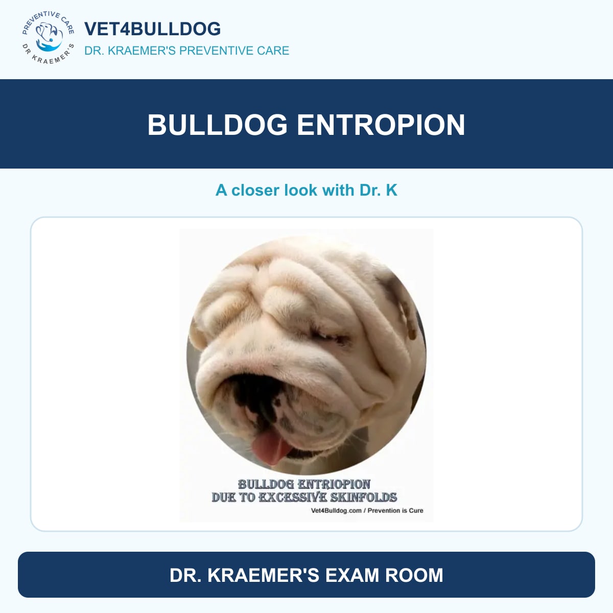
If entropion (inward-rolling eyelids) is present, a surgical correction is highly recommended
Entropion surgery helps to rotate the eyelids outward, reducing future distichia regrowth and corneal irritation.

#6. Supportive Topical Treatments
- Lubricants: To protect the cornea.
- Antibacterial Drops/Ointments: To prevent infection.
- Pain Relief: To reduce discomfort.
#7. Corneal Ulcer Healing
- Hyaluronic Acid Ointment
- Serum
- PRP (platelet-rich plasma) can promote faster healing.

#8. Pain Management
- NSAIDs: For inflammation and pain relief.
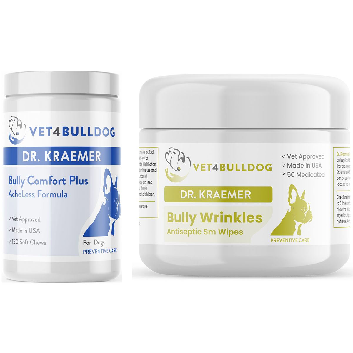
- Bull Therapeutic Supplements: Such as Dr. Kraemer’s V4B Bully Comfort & AchLess Formula Chews for long-term joint and eye comfort.
Bulldog Abnormal Eyelash and Corneal Ulcers TIPS & WARNINGS
Below are essential eyelid and eyelash tips and warnings courtesy of Dr. kraemer
#1: 🩺 Use an E-Collar Tip
- Keep a buster or e-collar in place until the abnormal eyelash is removed.
- Continue post-operatively, if needed, to prevent rubbing and pawing at the eye.

#2🩺 Eye Ophthalmic Topical Medicine Tip:
Use topical ophthalmic medications short-term to reduce pain, promote corneal healing, and combat bacteria.
#3 🩺Pain Relief Rx and Bulldog Supplements Tip:
Implement pain control prescriptions (RX) and consider supplements to aid in overall comfort and healing.
#4 🩺Thorough Eye Examination Tip:
- Always inspect the eyelids for abnormally ingrown eyelashes during any eye exam.
- Many abnormal eyelashes in Bulldogs are light-colored and thin, making them easy to miss.
- Use a magnifying headlight and view from different angles to ensure a complete evaluation.
In my exam room I always inspect bulldog’s eyelids with a magnifying head light from different angles.
#1⚠️ The Need for Eye Magnification & Illumination Warning:
- Bulldog distichiasis is difficult to detect with the naked eye.
- A magnifying glass and proper illumination are essential for accurate diagnosis.
#2 ⚠️Veterinary Doctor Error Warning:
Since distichiasis is breed-specific (bulldogs and French bulldogs), many veterinarians may fail to check for it or miss the condition entirely.
#3 ⚠️ Breeding Bulldogs and French Bulldog Warning:
Because abnormal eyelashes are hereditary, bulldogs with a history of eyelash disorders should not be bred.
#4 ⚠️ Potential Eyelid Scarring Warning:
Electrocautery and cryosurgery may lead to excessive scarring, potentially affecting eyelid function.
#5 ⚠️: Eyelash Regrowth & Repeat Procedures Warning:
Treated glands may still regrow abnormal hairs.
Other previously unaffected glands may start producing abnormal eyelashes later, requiring repeat procedures.
#6 ⚠️Ectopic Cilia Hidden Threat Warning:
Bulldog ectopic cilia (abnormal eyelashes growing from the inside of the eyelid) are:
- Extremely painful
- Difficult to detect (very short and deep inside the lid)
- Highly damaging to the cornea
Surgical removal is required ASAP to prevent severe corneal injury.
#7 ⚠️ Bulldog Entropion Complications Warning:
Bulldog entropion (inward-rolling eyelids) worsens distichiasis, increasing corneal trauma and discomfort.
#8 ⚠️ Risk of Eye Loss Warning:
Untreated, chronic bulldog distichiasis can result in:
- Severe corneal ulcers
- Infected, melting, or perforated ulcers
- Tragic loss of the eye if left untreated.
#9 ⚠️ Corneal Scarring & Vision Loss Warning:
Chronic corneal irritation from distichiasis or untreated ulcers can lead to:
- White opaque scars
- Fibrotic red scarring
- Black spots, cloudiness, and blood vessel formation over the cornea
- Permanent vision impairment
#10: ⚠️Eyes Rubbing & Itching, Other Causes Warning:
- Facial rubbing and itching can also be caused by:
- Bulldog allergies
- Bulldog and French bulldog skin fold dermatitis
Proper diagnosis is essential to help differentiate between eye irritation and allergy-related itching. To manage these conditions, consider:
- allergy control medications
- topical treatments
- Antiseptic skin fold wipes

Recommended by Owners Approved by Bulldogs
Your dog is not telling you how much this hurts
The single most useful thing I learned about Bulldog eyes is to stop using the dog’s behavior as the measure. In most breeds a corneal problem announces itself loudly: squinting, pawing, a dog who will not let you near the face. In this breed the surface of the cornea is less sensitive, so the same injury produces a fraction of the protest.
What you tend to get instead is subtle and visual rather than vocal. A film over the eye. More wetness on one side than the other. A dog holding his head slightly differently in bright light. Owners describe it as the eye just looking a bit off, and they are usually right, and they usually apologize for being vague. Vague is fine. Vague gets the dog seen.
This is also why I am firmer about eyes in Bulldogs than almost anything else on this site. A skin problem that waits a week is a skin problem that waits a week. The cornea is a few layers of tissue over the inside of the eye, and the distance between a surface scratch and something that threatens the eye itself is not very far. The judgment on how fast is your veterinarian’s, but the bias in this breed should be toward sooner.
And the test that answers the basic question is almost absurdly cheap. A drop of orange dye, a blue light, and an ulcer that was invisible a moment ago glows green. A painful eye in a Bulldog that has not been stained has not really been examined, and asking for it is a perfectly reasonable thing for an owner to do.
Plucking is a haircut, not a cure
Here is the distinction that decides whether your dog is back in a few weeks. Plucking pulls out the visible lash and leaves the follicle underneath entirely intact, so the lash regrows on its own schedule. It is quick, it needs no anesthetic, it gives immediate relief, and it is genuinely the right first move in plenty of cases. It is not a permanent answer and it is not usually sold as one, but owners hear the relief and reasonably assume the problem is solved.
The procedures that aim at the follicle itself are a different category. Freezing the follicle, or removing it surgically, are done under anesthesia with magnification, and they are what you reach for in the dog who keeps coming back. In a breed where anesthesia itself needs planning, the right conversation is not plucking against surgery in the abstract, it is how many times this has happened and what the eye has to lose.
The one I want owners to know by name is ectopic cilia, because it is the reason a case looks unexplained. That lash grows through the inner lining of the eyelid rather than along the margin, so it sits against the cornea with every blink and it is invisible from the outside. Finding it means everting the lid under magnification with the dog sedated. A Bulldog with repeated ulcers in the same eye and nothing obvious on the lid margin has earned that examination.
The last thing, and it is the one that costs owners most: the cone. A dog rubbing an eye with a healing ulcer can undo in one afternoon what a good few weeks of treatment achieved, and he will do it the moment the drops take the discomfort off. It is the most unpopular instruction in veterinary medicine and it is doing more work than most of the medication.
Let me be blunt, because eyes are not the place for soft edges. A corneal ulcer is treated with prescription medication and sometimes with surgery, and there is nothing on this site that touches it. If you are reading this with a cloudy or weeping eye in front of you, close the tab and telephone your veterinarian.
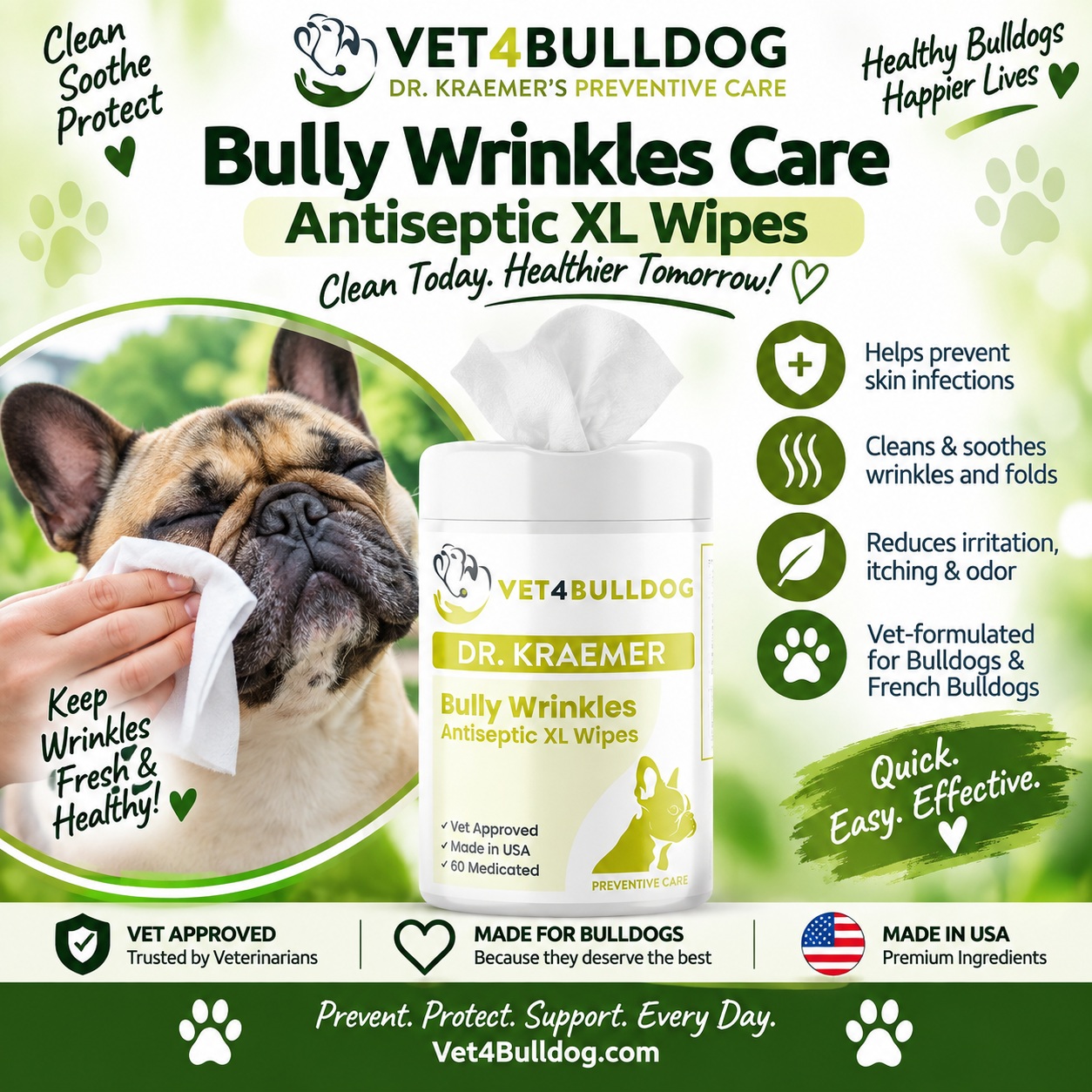
Where the products here have a genuine place is the part that comes before and after. Much of the hair that reaches a Bulldog’s cornea sits in the nose and facial folds rather than on the lid margin, and those folds also hold moisture and debris against skin that is already close to the eye. Antiseptic fold wipes and fold care are part of the multimodal approach to that skin and are used alongside what your veterinarian prescribes rather than instead of it. Keeping those folds clean and dry is ordinary maintenance, it is yours to do, and on a dog with a history of eye trouble it is worth doing properly.
Chronic weeping often leaves staining down the face as well, which is cosmetic rather than medical. The tear stain range deals with that side of it, and it is explicitly not treatment for whatever is causing the weeping.
To be clear: nothing here treats a corneal ulcer or an eyelash disorder. Topicals are used alongside veterinary treatment and are not a substitute for veterinary examination, diagnosis or treatment. This page is educational only.
How did I not notice? He never seemed to be in pain.
Because he was not giving you the signals you were watching for. Reduced corneal sensation is normal in short faced breeds, so the squinting and pawing that would make a different dog obvious are often simply absent. What you get instead is visual: cloudiness, a wetter eye on one side, a dog sitting with his back to the window. Owners who bring a Bulldog in saying only that the eye looks a bit funny are doing exactly the right thing, and the vagueness is the breed rather than the owner.
Does he really need the orange dye test?
It is the one test I would not skip. A drop of fluorescein under a blue light makes an ulcer visible that the naked eye cannot see, and it also tells you how deep and how wide it is, which changes the treatment. It takes about a minute, it does not hurt, and it is inexpensive. A red or painful eye that has not been stained has not been fully examined, and it is entirely reasonable to ask whether it has been done.
The lashes were plucked and the ulcer came back. Why?
Because plucking removes the lash and leaves the follicle, so it regrows. That is what plucking is, rather than anybody doing it badly. If your dog keeps relapsing, the two questions worth asking are whether the follicles have been destroyed rather than the lashes pulled, and whether the inner surface of the eyelids has been examined under magnification for an ectopic cilium, which is the lash that grows through the lid and cannot be seen from the outside. Repeated ulcers in the same eye with no clear cause is the classic picture for that.
I have leftover eye drops from last time. Can I start them?
Please do not, and this is the most important answer on this page. Some eye medications contain a steroid, and a steroid put onto an ulcerated cornea may deepen it rather than settle it, which is how a surface problem becomes a serious one. The bottle gives no warning and the drops look identical to the harmless kind. Leftover eye medication is one of the few genuine ways an owner trying to help makes things dramatically worse. Take the bottle to the appointment and let them read the label.
Does he have to wear the cone? He is miserable in it.
Yes, and I know exactly how that request lands. The reason it matters is the timing: the medication takes the discomfort away before the cornea has healed, so you get a comfortable dog with a vulnerable eye and an afternoon alone. One good rub against the carpet can cost you weeks of progress or the eye itself. If he genuinely cannot cope with the standard cone, ask about the softer and inflatable alternatives rather than abandoning it, because some dogs tolerate those far better and the point is only to keep the paw away from the face.
Will it keep happening, and does he need surgery?
Honestly, in this breed the underlying architecture does not change: the eye still sits forward in a shallow socket with hair nearby and a blink that does not fully close. So the goal is fewer episodes rather than a guarantee of none. What reduces the rate is dealing with the actual cause rather than each ulcer in turn, whether that is destroying the lash follicles, correcting an inward rolling lid, or managing the folds. Surgery is a conversation about how often this is happening and what the eye has to lose, and it is worth having with your veterinarian while the eye is still healthy rather than in the middle of a crisis.
Trusted by Vets ~ Recommended by Owners ~ Approved by Bulldogs
More Help from Dr. Kraemer
Eyes in this breed are a structural problem rather than an isolated one, so these are the pages I would read next.
- Entropion in Bulldogs and French Bulldogs
- Cherry Eye in Bulldogs and French Bulldogs
- Dry Eye and KCS in Bulldogs and French Bulldogs
- Nose and Face Skinfold Dermatitis
- Anesthesia in Bulldogs and French Bulldogs
- Brachycephalic Obstructive Airway Syndrome (BOAS)
About the Author
Roy Kraemer, DVM, is a veterinarian with extensive clinical experience caring for Bulldogs and French Bulldogs. His educational work emphasizes prevention, early recognition, appropriate at-home care, responsible medication use, and veterinary evaluation when disease is persistent or severe.