RE: Painfull red bulldog eye 🐶👁️
Good morning, Dr Kraemer; My 1.5-year-old Fr. bulldog Jussi seems to be squinting a lot, and I’ve noticed some rt eye redness, puffiness, discoloration, painfully squinting and she is trying to rub it. Should I come to see you for that🩺 ? What could be causing this, and what steps should I take for my bulldog’s eye discomfort?
Thank you!❤️#BulldogHealth #EyeIssues #EyePain
The elevated risk in bulldogs stems from their distinctive features, such as
➡️ flattened skulls
➡️ excess skin folds
➡️ prominent bulging eyes
➡️ Insufficient tear production
➡️ eyelid inward rotation
➡️ Ingrown or abnormal eyelashes
These factors contribute to a significantly increased susceptibility to corneal ulcers compared to most other dog breeds.

Bulldog Corneal Ulcer 5 X MUST KNOW
- Corneal ulcers are more common in bulldogs than in most dog breeds due to their flat faces and folds.
- In contrast to other dog breeds, bulldogs’ corneal ulcers are mostly due to dry eyes, abnormal eyelashes, and eyelids.
- Corneal ulcers can range from superficial and mild to deep and severe
- All bulldog and Fr. bulldog corneal ulcers are considered an emergency
- Prevention and care include Dr. Kraemer’s Bulldog Ophthalmic Bundles.
Members of this bulldog community prefer prevention over RX

Corneal Ulcer in Bulldogs and French Bulldogs: ANATOMY
Your bulldog’s cornea is the front part of the eye; it is transparent and acts like a windshield, covering the bulldog’s eye.
Its concave and clear nature makes it more noticeable when viewed from the side or in a profile position.
This is the area where we normally apply eye drops.
What Is the Purpose of Your Bulldog Cornea?
The cornea plays a vital role in your bulldog’s vision by allowing light to enter the eye and guiding it through the inner chambers and lens. This transparent structure directs light to the back of the eye, where the retina and optic nerve work together to create an image.
Think of the eye as a camera:
- The cornea is the lens
- The retina is the film capturing the picture
Just like scratches and “dirt spots” on a camera lens that can blur and reduce the quality of developed photos, scratches on the cornea and a decrease in transparency obstruct the passage of light.
If light cannot fully enter the eye, it will affect the sharpness of the image formed by the retina, directly impacting your bulldog’s eyesight.
BULLDOG CORNEA: 3 X LAYERS
The bulldog cornea is made from three specialized layers
- EPITHELIAL: the first layer, the outermost layer
- STROMAL: the second layer sandwiched in between the superficial epithelial and the posterior deep descement layer
- DESCEMET & ENDOTHLUM: the deepest, most posterior layer
BULLDOG TEAR PRODUCTION
The bulldog’s cornea is bathed and “lubricated” with tears.
The tear film helps provide nutrients and oxygen to the cornea.
Corneal Ulcer Types in Bulldogs and French Bulldogs
There are a few types of corneal ulcers, ranging from superficial and mild to deep and severe.
#1. BULLDOG CORNEAL EPITHELIAL ULCER 🚨
- It can be difficult to see with the naked eye and might require a stain for a diagnosis
- It is considered the least severe
- And typically responds promptly to appropriate treatment, resulting in quick healing.
The superficial corneal ulcer in bulldogs is the most prevalent type
#2. BULLDOG STROMAL ULCER 🚨
A bulldog stromal corneal ulcer is
- deeper
- easier to see with the naked eye
- It’s usually cloudy-looking
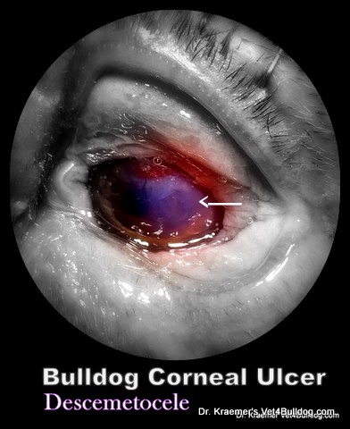
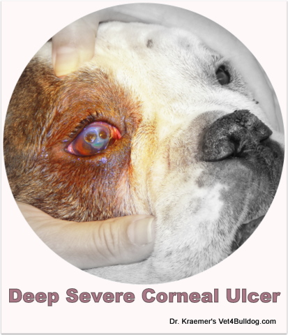
#3. BULLDOG CORNEAL DESCEMET ULCER 🚨
Bulldog descemtocele ulcer is the
- deepest
- most serious
- can melt and perforate
- challenging to treat
- duration to full healing is often much longer
🚨A Descemtocele deep corneal ulcer has the potential to perforate, leading to permanent loss of sight in your bulldog. Thus, it requires urgent care.
What Causes Corneal Ulcers in Bulldogs and French Bulldogs?
For most breeds, exterior trauma is the #1 cause; it is not so for bulldogs
In contrast to other breeds, the most common causes of bulldog corneal ulcers are due to the breed’s anatomical characteristics and structural defects:
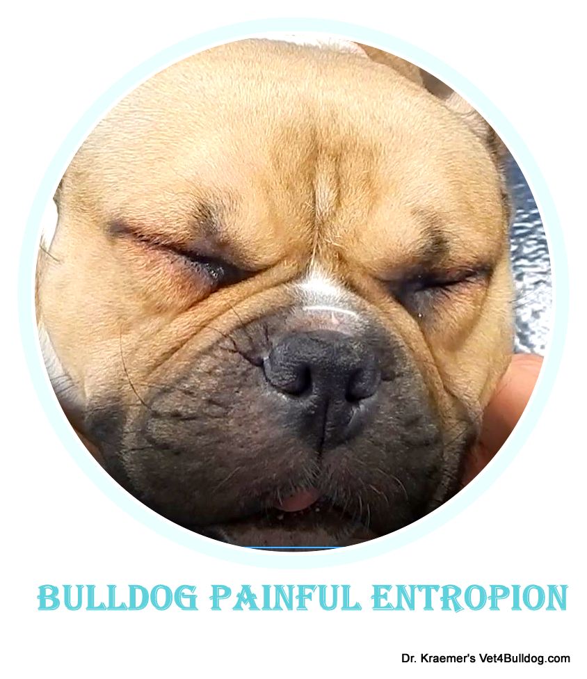
#1. ULCER DUE TO ENTROPION
The inversion of eyelids, caused by the abundance of facial skin folds, leads to the lid and eyelashes rubbing against the cornea, causing irritation and injury.

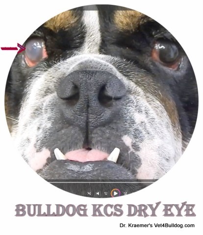
#2. CORNEAL ULCER DUE TO DRY EYE
Bulldog dry eye (keratoconjunctivitis sicca KCS ) is a prevalent immune-mediated condition unique to the breed that impacts tear production.
The deficiency in tears deprives the cornea of
- vital nutrients
- oxygen
- lubrication,
– This leads to chronic corneal disease and ulceration.
#3. CORNEAL ULCER DUE TO ABNORMAL EYELASHES
Irregular eyelashes that come into contact with the cornea can cause pain and an urge to rub, leading to direct and indirect injuries to the cornea.
Direct injuries happen when the eyelashes touch the cornea, while indirect injuries result from rubbing and itching in response to the irritant.
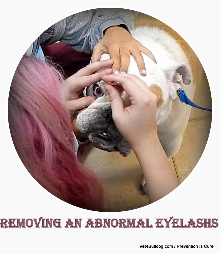
Examples of abnormal eyelashes include:
- distichiasis
- trichiasis
- ectopic cilia
#4. BULLDOG CORNEA ULCER DUE TO TRAUMA
While less frequent, corneal ulcers may also arise from external traumatic incidents, such as:
- Running into a rose bush
- Cat Scratch
- A chemical injury, such as soap or shampoo
- Accidentally rubbing the eye due to allergies such as bulldog food allergy and atopic dermatitis
- Rubbing the eyes due to bulldog skinfold dermatitis

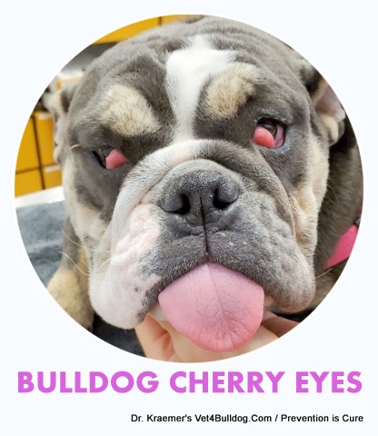
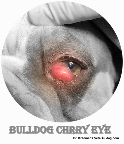
#5. BULLDOG CORNEAL ULCER DUE TO CHERRY EYE
Cherry eye is common in French bulldog and English bulldog puppies.
A large, protruded cherry eye could rub the cornea and also impede tear production
#6. CORNEAL ULCER DUE TO EXPOSURE KERATOPATHY
Injuries resulting from exposure are a consequence of the breed’s brachycephalic confirmation. The compressed skull affects the eye sockets, causing bulging eyes and an inability of the eyelids to close fully.
These bulging eyes, coupled with insufficient tear distribution, contribute to corneal exposure and issues with the tear film
#7. CORNEAL ULCER CAUSE: OTHER
➡️ CORNEAL ULCER: HORMONAL
Endocrine diseases such as
- diabetes
- hypothyroid
- Cushing’s
➡️ VIRAL & BACTERIA CORNEA ULCER
➡️ CORNEAL DYSTROPHY
➡️ FACIAL NERVE PARALYSIS:
can contribute to dry eyes by disrupting the blinking mechanism, consequently affecting the spread of tear film.
Insufficient tear film distribution leads to dry eyes, and persistent dry eye conditions can result in corneal ulcers.
Corneal Ulcers in Bulldogs and French Bulldogs PRESENTATION
Depending on severity, your bulldog may exhibit a wide range of symptoms
1. CORNEA ULCER SEVERE PAIN
Pain is typically manifested by:
- Blinking & Squinting
- Tearing & Discharge
- Rubbing & Pawing
- Shut often swollen eyelids (closed eye)
- Other: Change in appetite and other behavioral abnormalities associated with PAIN
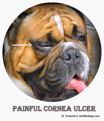
Bulldog and French bulldog corneal ulcers can be very painful
2. CORNEA ULCER DISCOLORATION
- white patches
- opaque area
- vascular areas
- pigmented areas
- redness
- hemorrhagic
3. CORNEA ULCER PUNCTURE
Visible “holes,” punctures, and concave lesions are prone to infection and can rapidly deteriorate. It should always be regarded as an emergency.
Corneal Ulcer in Bulldogs and French Bulldogs: DIAGNOSIS
A. DOCTOR EXAM
Visual examination and ophthalmoscopy exam:
B. EXHIBITING PAIN
Evidence of ocular discomfort, squinting, abnormal blinking,
C. ITCHING AND RUBBING
Evidence of skin irritation and redness that can trigger rubbing and pawing
Common cause
D. BULLDOG ENTROPION
Evidence of underlying traumatic causes, such as abnormally rotating eyelids.
E. ABNORMAL EYELASHES:
It usually requires a magnifier and special bright illumination.
H. OTHER BULLDOG OCULAR DISEASES:
We are looking for evidence of ocular diseases such as glaucoma and uveitis, including ocular discoloration and bulging consistently.
The diagnosis will require tonometry (measurement of the intraocular pressure)

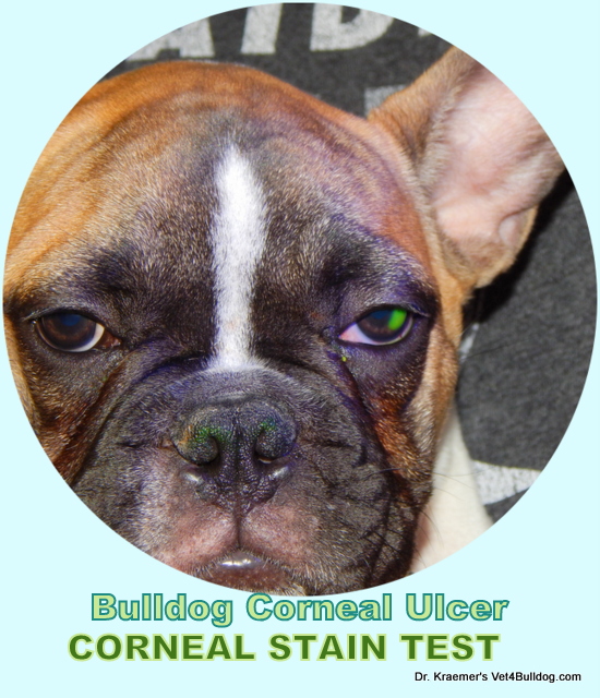
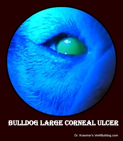
BULLDOG CORNEAL ULCER STAINING
Using a special corneal stain and ultraviolet light, we can detect ulcers that might be difficult to see on an exam.
The fluorescein stain test is the most common bulldog cornea ulcer test performed
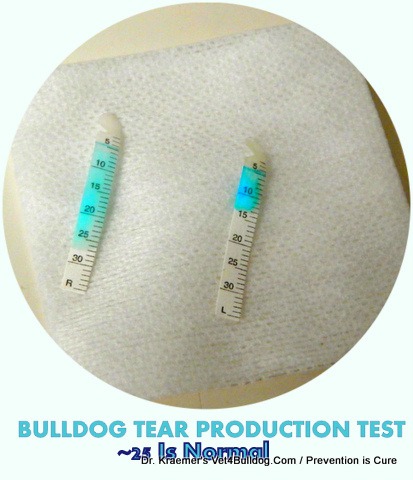
1. SCHIRMER TEAR TEST
A tear production test should be done on any bulldog presented with a corneal ulcer.
2. CORNEA ULCER CULTURE & CYTOLOGY
A deeper, more advanced corneal ulcer or a non-healing one will require a sample for culture and cytology.
How to Treat Corneal Ulcers in Bulldogs and French Bulldogs?
Treatment depends on the type of ulcer and the underlying cause
TOPICAL TREATMENT
1. TOPICAL ANTIBIOTIC: ophthalmic antibacterial ointment or drops placed a few times daily
2. TOPICAL SPASM & PAIN RELIEF: Ophthalmic atropine drops or ointment can help with ocular pain and discomfort
3. LUBRICATION AND TEAR PRODUCTION: Ophthalmic drops or ointment should be used for excessive exposure.
Examples are:
- saline
- hyaluronic acid
lifelong drops/ointment to help maintain adequate tear production
4. SERUM & PRP: Autologous serum eye drops and PRP (platelet-rich plasma) can also help heal some types of corneal ulcers
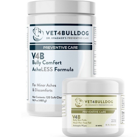
5. BULLY SKINFOLD ANTISEPTIC WIPES:
It is important to recognize the cause and effect of itching, painful nasal or facial bulldog skinfold dermatitis, and bulldog corneal ulcers. The proximity of the infected folds to the eyes means that an itching, pawing, or rubbing of the infected skinfold can lead to an accidentally injured cornea.
Preventive care for your bulldog’s skinfold dermatitis can prevent a more serious and costly corneal injury.

Using Dr. Kraemer’s V4B Antiseptic Bully Skinfold XL wipes is the ideal preventive and maintenance therapeutic remedy for this common bulldog condition
6. SYSTEMIC RX
- Pain Relief: Oral NSAID anti-inflammatory and pain relief analgesics might be needed, especially for bulldogs with deep stromal and descemetocele corneal ulcers
- Antibiotics: oral antibiotics are not necessary for superficial ulcers but often are for deep stromal and/or descemetocele corneal ulcers
Corneal Ulcers in Bulldogs and French Bulldogs: SURGERY
1. GRID KERATECTOMY:
Surgery might be necessary for indolent ulcers to help remove detached or poorly healing corneal epithelium
2. CORNEAL GRAFT:
A graft might be required to help save a corneal descemetocele (deep melting ulcer)
3. ENTROPION SURGERY:
When your bulldog’s inward eyelid rotation is the source of the corneal ulcer, entropion surgical treatment is immediately required.
4. TEMPORARY ENTROPION SURGERY:
Bulldogs experiencing inwardly rotating eyelids (entropion) can find relief through this procedure. The surgery is also advantageous for eyelids experiencing spasms due to pain. Moreover, it serves as an emergency measure until a permanent entropion correction can be scheduled.

It is usually done with surgical staples
This procedure can be swiftly performed in an emergency setting within seconds and without anesthesia, or with mild sedation if necessary.
5. ABNORMAL EYELASH SURGERY:
Abnormally ingrown eyelashes that are either growing in the wrong direction or inside the eyelid will require surgical intervention.
It is usually done with liquid nitrogen freezing (cryo freezing)

5. CHERRY EYE SURGERY:
Some protruding third eyelid glands (“cherry”) can rub against the cornea, resulting in injury.
Bulldog cherry eye can also hinder tear production, leading to dry eyes and subsequently causing corneal ulcers.
Cherry eye surgical repair must preserve the glands; they are critical for tear production.

Bulldog and French Bulldog Corneal Ulcer TIPS & WARNINGS
Below are selected corneal ulcer tips and warnings courtesy of Dr. Kraemer
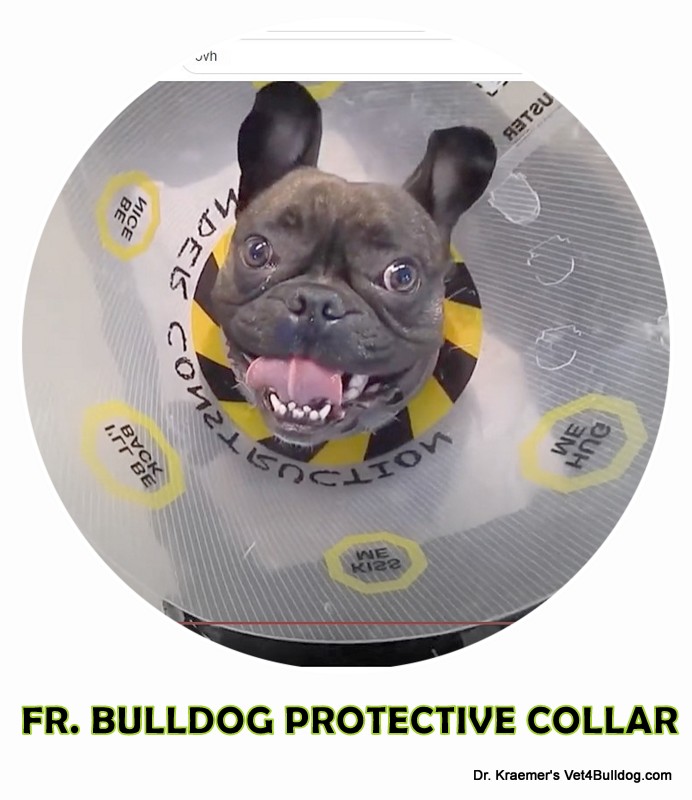
#1 🩺 PROTECTIVE GEAR TIP:
Place a buster collar to prevent any further injury
#2 🩺 R/C EXAM TIP:
r/c exams are critical to confirm healing; they usually require a cornea-negative stain
#3 🩺 OPHTHALMOLOGIST TIP:
A referral to a specialist might be required for a nonhealing corneal ulcer or a severe, deep, melting, or perforating one.
#1 ⚠️ ATROPINE WARNING:
The atropine ophthalmic drops will dilate your bulldog’s pupils in addition to alleviating the spasm. As a result, you should avoid exposing your bulldog to harsh light.
#2 ⚠️ STEROID WARNING:
Topical ophthalmic steroids are widely used to treat various eye conditions, but they must only be applied after a corneal ulcer has fully healed.
It is vital to NEVER use steroids on an active corneal ulcer, as they can hinder healing and potentially cause severe complications.
If, after using any topical ophthalmic containing a steroid (indicated by terms like “pred,” “dex,” or “hydro,” your bulldog symptoms get worse, such as
- squinting
- abnormal blinking
- discolored cornea
- discomfort
-in that case, you must discontinue the topical ophthalmic application and seek veterinary attention.
#3 ⚠️ EMERGENCY WARNING:
Corneal ulcers constitute an emergency. If you observe your bulldog squinting or notice the cornea appearing discolored, vascular, or opaque, it is imperative to seek veterinary assistance promptly.
Recommended by Owners Approved by Bulldogs