Subject: Concern About New Skin Mass 🐾🚨
Dear Dr. Kraemer,
I hope this email finds you well. I’m writing to you because I’ve noticed a new skin mass on my 9-year-old French Bulldog, Franki and it’s causing me quite a bit of worry. 🐶😟 Given the prevalence of tumors and cancer in bulldogs, I’m extremely concerned about what this could mean for her health.🙏💕
Warm regards,
Tim and Bella 🙏💕 #BulldogHealth #WorriedDogMom #petcancer
In my practice, I encounter a diverse array of bulldog tumors. Unfortunately, many pet owners mistakenly equate a tumor with cancer, leading them to anticipate dire consequences. However, it’s important to note that a significant portion of these tumors, growths, and masses are benign. In some cases, they may be localized and slow-growing, allowing for safe monitoring or surgical excision with minimal long-term effects.
Conversely, while some tumors may initially seem small, benign, and insignificant, leaving them untreated can lead to rapid progression, with the potential for aggressive behavior and deadly consequences.
Bulldog Cancer and Tumors 5 X MUST KNOW
- Not all tumors are cancerous and malignant; many are benign
- They are more common in older bulldogs; they can be external or internal.
- Diagnosis and prognosis should be based on testing, not visual inspection
- Treatment depends on age, cost, and quality of life merits.
- Prevention includes early detection, lifestyle, and Dr. Kraemer’s Immune Bundles
Wellcome to “Prevention Over RX” bulldog community

It is crucial not to jump to conclusions or overlook any lumps, masses, or growths
It’s advisable to consult your veterinarian promptly and have them examined.
Tumors-Growth-Cancer in Bulldogs MYTH and FACT:
Listed below are some of the most common tumor myths
PAINLESS TUMOR MYTH:
MYTH: “ My bulldog’s recently found tumor does not seem to inflict any pain, it is not bothering her one bit, thus it can be ignored.”.
FACT: Pain or its absence should not be the sole criterion for assessing tumors. It’s essential to recognize that malignant tumors can sometimes be painless, while benign tumors may cause discomfort. Therefore, relying solely on pain as an indicator can be misleading.
It’s important to promptly consult with a veterinarian for a thorough evaluation and proper diagnosis of any suspected tumors, regardless of the presence or absence of pain.
PAINFUL TUMOR MYTH:
MYTH: “My bulldog’s recently found tumor seems to inflict pain. Every time I touch it, she flinches, thus it must be cancerous.”
FACT: Painful growths, masses, and tumors do not always indicate malignancy, and their presence alone is not a reliable indicator of severity. Pain can result from various factors such as pressure, infection, necrosis, or an allergic reaction, as seen with mast cell tumors.
Therefore, it’s crucial not to solely rely on pain as a diagnostic criterion and to consider a range of potential causes when assessing such symptoms. Consulting with a veterinarian for a comprehensive evaluation and diagnosis is essential for appropriate management and treatment.
BULLDOG TUMOR SIZE:
MYTH: “Dr. Kraemer, my French bulldog skin tumor is only pea size, thus can be ignored.”
FACT: Do not make assumptions or overlook even small lumps, masses, or growths. The size of a tumor should not dictate your course of action.
It’s crucial to consult your veterinarian and have it thoroughly examined. While small mast cell tumors can become aggressive if left untreated, large lipomas (fatty tumors) are often harmless. Only a professional evaluation can provide an accurate diagnosis and guidance on appropriate measures.
BULLDOG TUMOR DIAGNOSIS:
MYTH: “Dr. Kraemer, you have seen all kinds of tumors, and we heard that you are a bulldog expert. If I send you a photo of my English bulldog’s new skin tumor, would you be able to tell me if Rambo will be okay?”
FACT: Your veterinarian can conduct an initial evaluation of your suspected tumor based on factors such as:
- location
- size
- color
- consistency
- mobility
However, without high-power microscopic vision, a definitive diagnosis may be challenging.
Therefore, it’s typically recommended to perform either cytology or a biopsy (histopathology) for further evaluation, treatment options, and prognosis. These diagnostic procedures allow for a more accurate assessment of the tumor’s nature, helping to guide appropriate treatment decisions.
BULLDOG TUMOR VISUAL EXAM
MYTH: “Dr. Kraemer, you have seen all kinds of tumors, and we heard that you are a bulldog expert”, If I send you a photo of my English bulldog’s new skin tumor, would you be able to tell me if Rambo will be ok?”
FACT: Your veterinarian can make an initial evaluation based on the location, size, color, consistency, and mobility of your bulldog’s suspected tumor, but he or she does not possess high-power microscopic vision.
Visual inspection is not adequate, other tests are required, such as cytology and biopsy (histopathology), before treatment options, and prognosis are discussed.
Bulldog Tumors-Growth-Cancer Types:
- Benign vs. Metastatic cancerous types
- Local vs. diffuse
- Focal vs. Multifocal
- Lobar vs multilobar
- Necrotizing and Infected
- Slow-growing vs Locally aggressive
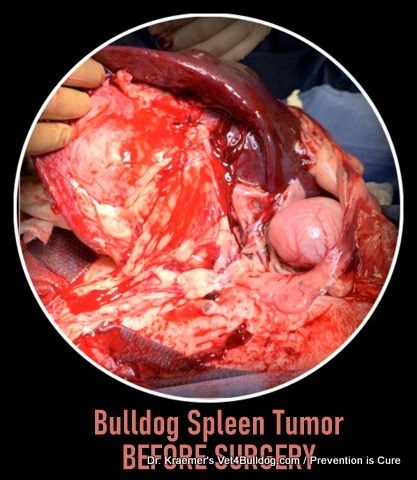
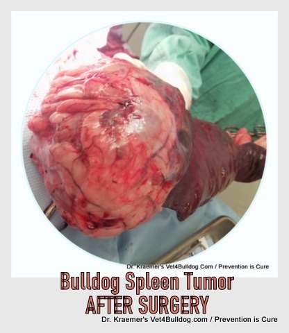
- Bleeding: Bleeding is often the result of spleen or liver tumors and ones originating from blood vessels (“hemangio” types)
- Secreting: common to secreting glands like the pituitary, thyroid, and adrenal
Tumor and Cancer in Bulldogs Clinical Presentation Examples:
- BRAIN OR SPINAL TUMOR: neurological deficits and abnormalities
- SPLEEN TUMOR: a bleeding tumor that is causing
- leathery
- labored breathing
- pale gums
- GI TUMOR:
- weight loss
- vomiting
- diarrhea
- abdominal pain
- distended, hard abdomen
- HEART-BASED TUMOR:
- distended abdomen due to right-sided heart failure
- heart murmurs
- heart irregularities
- cough and labor breathing
Bulldog Cancer Test Results Examples:
- BLEEDING TUMORS: low red blood cell on a CBC blood test
- BONE MARROW TUMOR: abnormal cells on a CBC cytology
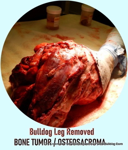
- BONE TUMOR: bone fractures and osteolytic periosteum on radiographs
- HEART BASE TUMOR: seen on an ultrasound and arrhythmia on ECG
Cancer and Tumors in Bulldogs / TREATMENT OPTIONS:
It’s essential to engage in open and honest discussions with both the caregiving team and family regarding treatment options.
Making decisions in this context can be emotionally taxing, and filled with heartache and stress.
Ensure that the medical team does not pressure you into making a decision.
You should never be made to feel guilty or perceived as a “bad owner” by the medical team.
If necessary, do not hesitate to politely refuse and seek a caregiver who offers a treatment plan tailored specifically to your needs and preferences.
When choosing a treatment, consider the following factors:
- Age and Time: Will the treatment significantly extend the life of my aging pet?
- Quality: Will the treatment improve my pet’s quality of life, considering potential issues like pain, nausea, diarrhea, incontinence, etc
- Side Effects: What kind of side effects might my bulldog and my family need to manage?
- Cost: Be mindful that many treatments can be expensive.
- Commitment: Consider the time commitment required for the chosen treatment.
It’s important to make choices that you feel comfortable and confident with.
Tumors-Growth-Cancer in Bulldogs and French Bulldogs TIPS & WARNINGS:
Below are a few selected tumor tips and warnings, courtesy of Dr. Kraemer

BULLDOG CANCER TESTING TIP:
Although blood tests may not directly diagnose tumors in many cases, they can provide valuable information about organ function, inflammation, and potential abnormalities that may indicate underlying health issues.

Additionally, certain tumors affecting the bone marrow and blood cell production may be detectable through specific blood tests. Therefore, including blood tests as part of the diagnostic workup can aid in identifying these types of tumors and guide further evaluation and treatment.
TUMOR IMAGING DIAGNOSIS TIP:
- Radiographs
- Ultrasound
- CT
- MRI
Screening for tumors and cancer often involves radiographic imaging and, in some cases, additional modalities such as ultrasound, computed tomography (CT), and magnetic resonance imaging (MRI). These imaging techniques are typically recommended to detect metastatic disease, which refers to the spread of the tumor to other parts of the body.
RADIOGRAPH
Radiographs (X-rays) are commonly used to evaluate the presence of tumors and assess their size, location, and potential impact on surrounding structures. However, they may not always provide sufficient detail to detect metastases or evaluate soft tissue abnormalities comprehensively.
ULTRASOUND
Ultrasound imaging utilizes sound waves to create detailed images of internal structures, allowing for the visualization of tumors and their characteristics. It is particularly useful for evaluating abdominal and soft tissue tumors.
CT SCAN
CT scans provide cross-sectional images of the body, offering detailed views of internal organs and structures. They are valuable for detecting small tumors, assessing their extent, and identifying metastases, especially in areas that may be challenging to evaluate with radiographs alone.
MRI
MRI utilizes magnetic fields and radio waves to generate detailed images of soft tissues, providing high-resolution views of tumors and surrounding structures. It is particularly useful for evaluating tumors in the brain, spinal cord, and soft tissues.
By utilizing a combination of these imaging modalities, veterinarians can effectively screen for tumors, assess their characteristics, and detect any signs of metastatic spread, enabling more accurate diagnosis and treatment planning.
BULLDOG CANCER TREATMENT TIP:
Treatment for tumors may involve various modalities, including surgery, chemotherapy, and radiation therapy, and be tailored to each case, considering factors such as:
- pet’s age
- financial considerations
- potential adverse effects,
- preferences of the pet owners
These factors are crucial in determining the best course of action for achieving the optimal outcome.

However, despite the individualized nature of treatment decisions, several common considerations should always be discussed:
- Nutrition: Providing a proper diet rich in essential nutrients is vital for supporting overall health and potentially aiding in the treatment process.
- Calm Environment: Minimizing stress and anxiety in the pet’s environment can promote well-being and potentially enhance the effectiveness of treatment.
- Comfort: Palliative pain control measures should be implemented to ensure the pet’s comfort and quality of life, especially if the tumor is causing discomfort.
- Alternative and complementary therapies, such as supplements and nutraceuticals, can play a supportive role in the treatment of tumors in pets. While these therapies are not typically used as standalone treatments, they may complement conventional approaches and contribute to the overall well-being of the pet.
By addressing these common considerations in addition to tailoring specific treatments to the individual needs of the pet, we strive to achieve the best possible outcome in the management of tumors. Supplementation typically includes therapeutic that help
- replenish the gut microbiome
- soothe gastrointestinal irritation
- detoxify the liver toxicity
- boost the immune system
- relax and calm
- essential vitamins & minerals

Bulldog Cancer PREVENT & CARE:
Regardless of your treatment, care, and therapy choices, taking a holistic approach to treatment is essential for achieving the best possible results. It’s important to prioritize a nutritious diet, therapeutic supplementation, pain control, and a relaxing environment free from stress.
Bulldog Immune Support & Anti-Oxidants

Bulldog Stress Relief & Anti-Anxiety

Bulldog GI Health

Bulldog Comort & Achless

BULLDOG CANCER DIAGNOSIS WARNING:
Not all tumors are externally visible or palpable; some may be located internally within your pet’s skeletal system, deep tissue, abdominal or chest cavity, brain, or internal organs. These types of tumors are often identified through imaging techniques such as radiographs (X-rays), ultrasound, magnetic resonance imaging (MRI), and computed tomography (CT).
However, while imaging can provide valuable information about the size, location, and characteristics of internal tumors, a definitive diagnosis typically requires a biopsy or cytology. These procedures involve obtaining a tissue sample from the tumor for microscopic examination,
Tumors-Growth-Cancer in Bulldogs and French Bulldogs MOST COMMON:
In bulldogs, like many other dog breeds, various types of tumors can occur. Here are some of the most common ones seen in the breed:
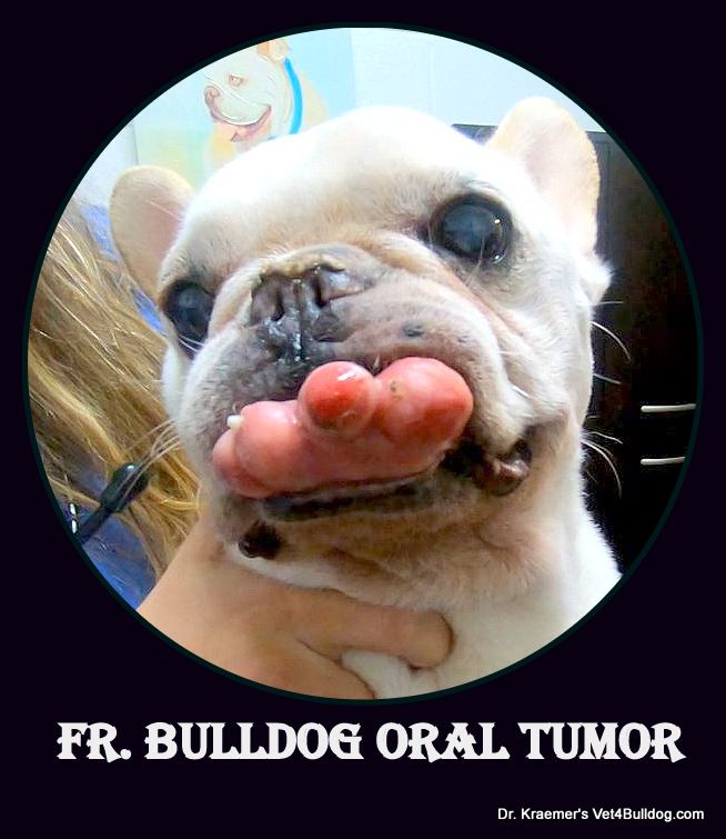
BULLDOG MAST CELL TUMOR
Bulldog mast cell tumors (MCTs) are clusters of mast cells that can lead to harmful effects. They may manifest as itchy lesions and can remain small and inconspicuous-looking for extended periods, sometimes months or even years. Due to histamine-induced gastric acid secretion triggered by MCTs. bulldogs may exhibit gastrointestinal symptoms, such as
- irritation
- vomiting
- diarrhea
- weight loss
- bloody stool
The appearance of mast cell tumors in bulldogs can vary greatly and may resemble lipomas, skin tags, or insect bites. Therefore, it’s crucial to perform a biopsy or cytological examination to confirm the diagnosis and differentiate MCTs from other benign conditions.
To prevent the transformation of MCTs into a more aggressive form, it’s generally recommended to surgically remove mast cell tumors in bulldogs and French bulldogs. Early detection and intervention can improve the prognosis and reduce the risk of complications associated with MCTs.
Regular monitoring and prompt veterinary attention are essential for managing these tumors effectively.
BULLDOG MAMMARY GLAND BREAST TUMOR
Bulldog mammary gland tumors (MGT) are tumors that originate from the breast tissues. Approximately 50 percent of these tumors are malignant, but spaying your bulldog or French bulldog before their first estrus cycle (heat cycle) can reduce the chances of MGT by over 95%.
If a suspect tumor is identified, it should be biopsied and removed along with any associated breast tissue. Pre-surgical chest radiographs are often performed to rule out any spreading of the tumor to the lungs.

If your bulldog or French bulldog has not been spayed, it is recommended to consider spaying at the same time as tumor removal surgery.
Spaying not only reduces the risk of MGT but also eliminates the possibility of future hormone-related reproductive health issues.
BULLDOG and FRENCH BULLDOG LIPOMA
Bulldog lipomas, commonly known as fatty tumors, are frequently observed in older bulldogs and French bulldogs. These tumors tend to grow slowly and are typically soft to the touch, located beneath the skin layer.
Infiltrative lipomas are a less common variant that is less well-defined and usually situated deeper within the tissues.
Due to the benign nature of most bulldog and French bulldog lipomas, removal is generally not necessary. However, if they grow excessively large and begin to impact your pet’s comfort or mobility, or if they are of the infiltrative type, surgical removal may be recommended.
Regular monitoring of lipomas by a veterinarian is important to assess any changes in size, texture, or appearance, and to determine if intervention is necessary for your pet’s well-being.
BULLDOG HEART-BASED TUMOR
Bulldog heart base tumors, such as aortic body tumors (e.g., chemodectoma), are more commonly observed in brachycephalic breeds like English bulldogs and French bulldogs.
These tumors are often nonresectable, meaning they cannot be surgically removed, but they tend to grow slowly and are unlikely to metastasize (spread to other parts of the body).
In cases where there is significant pericardial effusion (fluid around the heart) associated with these tumors, a procedure called pericardiectomy may be performed as a palliative measure. Pericardiectomy involves surgically removing a portion of the pericardium (the sac around the heart) to relieve pressure and improve cardiac function.
While bulldog heart base tumors present challenges in terms of treatment options, managing symptoms and providing palliative care can help improve the quality of life for affected dogs. Regular monitoring by a veterinarian is essential to assess the progression of the tumor and adjust treatment as needed.
Bulldog Growths Cancer and Tumors / OTHER
- Soft Tissue Sarcomas: Soft tissue sarcomas can develop in the muscles, connective tissues, or other soft tissues of the body. They often present as firm, non-painful masses beneath the skin.
- Melanomas: Melanomas are tumors that arise from melanocytes, the pigment-producing cells. They can occur on the skin or in mucous membranes and can be benign or malignant.
- Osteosarcomas: Osteosarcomas are aggressive bone tumors that commonly affect large and giant breeds, like English. bulldogs. They typically occur in the limbs, can cause lameness and pain, and carry a poor prognosis
- Lymphomas: Lymphomas are cancers of the lymphatic system and can affect various organs and tissues, including lymph nodes, spleen, liver, and bone marrow.
- Histiocytomas: These are benign tumors derived from histiocytes, a type of immune cell. They typically occur as solitary, raised nodules on the skin of young dogs.
- Fibrosarcomas: Fibrosarcomas are malignant tumors arising from fibrous connective tissue. They can occur in various locations, including the skin, subcutaneous tissues, and bones.
- Sebaceous Gland Tumors: These tumors originate from the sebaceous glands in the skin and can vary in appearance and behavior. They may appear as raised, firm nodules on the skin.
- Hemangiosarcomas: These are malignant tumors derived from blood vessel cells. They can occur in various organs, including the
- skin
- spleen
- liver
Recommended by Owners Approved by Bulldogs